2026.03 ECVD HistoRound — Group 1「Dermal reaction patterns」(Monika Welle)⚠️ 只有最後 23 分鐘
官方標題:Group 1 MW March dermal reaction patterns。 ⚠️ 官方上傳的影片只有 23 分鐘,而且從中段開始(開頭是「這張不在清單上…我們看 B1255」;Q&A 提到「前面看過的 musculopathy/elastosis」都不在片中)。前面的真皮反應型態(膠原、彈性纖維等細胞外基質變化)沒有錄到。Monika 說 4 月接著講真皮的 depositions(但官方清單沒有 4 月 Group 1 場次,下一場是 5/7 Perivascular)。 逐字稿:
逐字稿/2026.03 官方 Group 1 March 逐字稿(英文原文,僅後 23 分鐘).txt;截圖:attachments/2026.03_official/。
⭐ 本場考點速記(30 秒複習)
- 看到寄生蟲一定要量大小+描述構造;沒有尺就用淋巴球(8–9 µm)當比例尺(Monika:“if you see a parasite you should try to at least measure it somehow in size”)。
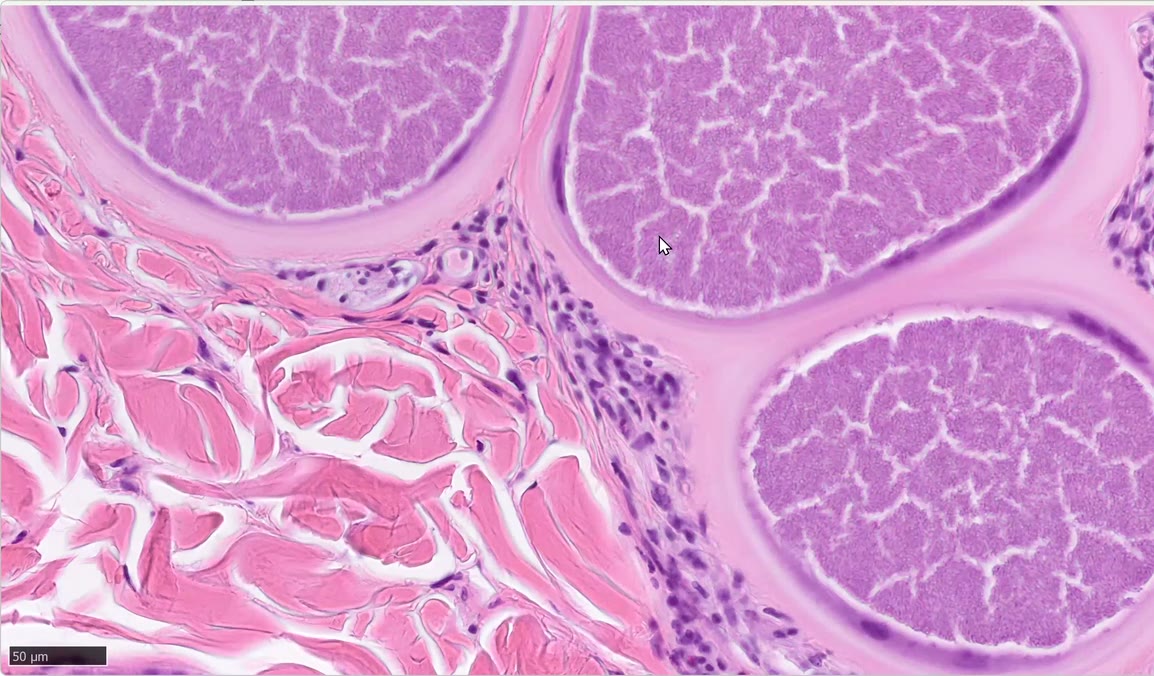
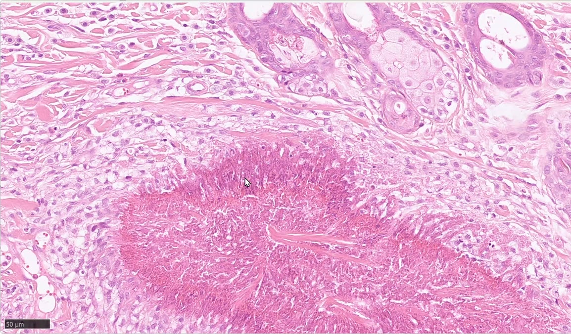
- Besnoitia 囊 200–800 µm(“there’s no other parasite cysts that it has this size”):淡嗜伊紅環=壓扁的真皮結締組織 → 壓扁的宿主細胞 → 數百個香蕉形 bradyzoites(約 5–7 µm)。
- 約 50% 真皮被取代時要寫 abundant,不是 multiple;牛的深層叫 hypodermis,不叫 panniculus。
- Eosinophilic plaque vs(collagenolytic)eosinophilic granuloma = flame figure 周圍有沒有 epithelioid macrophages/multinucleated giant cells 圍住;spongiosis 不能拿來區分。
- Palisading 這個詞保留給組織球;嗜伊紅球在病灶邊緣堆積只是病灶長大的樣子。
- Solar elastosis 的彈性纖維=Walder’s spaghetti:細而長(通常不是藍色)。
- 已複習
📋 本段案例(2 片+Q&A)
| # | 玻片 | ⏱ | 物種 | 診斷 | 一句話重點 |
|---|---|---|---|---|---|
| 1 | B1255 | 00:00–09:30 | 牛 | 真皮內 Besnoitia 囊(besnoitiosis) | 寄生蟲一定要量大小;200–800 µm、囊壁=壓扁的膠原+宿主細胞、內含香蕉形 bradyzoites |
| 2 | B32 | 10:50–20:30 | 貓 | Eosinophilic plaque → 正在轉成 collagenolytic granuloma | flame figures;有 epithelioid macrophages/giant cells 圍住才叫 granuloma |
| Q&A | — | 20:59–22:30 | — | Elastosis:Walder’s spaghetti | 細、長、(藍書說)偏藍的細絲 |
🧫 1. 牛真皮 Besnoitia 囊(B1255,⏱ 00:00–09:30)

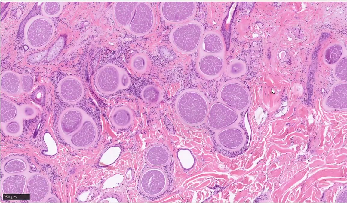
學員(Abigail)描述: 牛頭部皮膚(simple follicles、大腔汗腺);低倍下多發卵圓形囊狀構造、周圍嗜伊紅環,以淺真皮為主、局部延伸到深部;囊周圍有發炎細胞(histiocytes、eosinophils)。
Monika 的修正:
- 牛的深層不叫 panniculus,叫 hypodermis(anagen 毛球所在處以下就是 hypodermis)。
- 「multiple」→ 應寫 abundant——約 50% 的真皮被囊取代。
- ⭐ 最重要的是把囊描述清楚:大小+外觀。 沒尺可用淋巴球(8–9 µm)當比例尺;本例約 200–800 µm(文獻數值)——沒有其他寄生蟲囊這麼大。
- 囊的構造(由外而內):淡嗜伊紅環=被壓扁的真皮結締組織 → 被壓扁的宿主細胞(含胞漿) → 內含數百個香蕉形 bradyzoites(約 5–7 µm,比淋巴球略小)。
- 發炎相對輕微:因為蟲在細胞內、在真皮。
- 發炎細胞:多為淋巴球,另有嗜伊紅球與組織球。
- 診斷:Besnoitia(牛 besnoitiosis)——當作「真皮內的寄生蟲囊」示範。
🔬 Morph/診斷(依逐字稿補)
- Morph(morphologic description,型態描述;依學員描述+Monika 修正整理):Superficial dermis, extending to hypodermis: abundant (≈50% of dermis replaced) large oval intracellular parasitic cysts (≈200–800 µm), with a pale eosinophilic wall of compressed fibrous connective tissue, a compressed host cell with cytoplasm, and hundreds of banana-shaped bradyzoites; mild inflammation of lymphocytes, eosinophils and histiocytes.
- 診斷:Bovine besnoitiosis(Besnoitia 囊)。
🎯 教學重點(依逐字稿補)
- 囊是這張片子「最重要的東西」(“this is the most important thing you should describe them a little bit better in terms of size and how they look like”)→ 描述要有數量用詞(abundant)+大小+由外而內的構造。
- 大小本身就幾乎是診斷:沒有其他寄生蟲囊有這麼大。
- 學員把淡嗜伊紅環只想成「fibrosis」→ Monika 澄清是被壓扁的纖維結締組織(compressed fibrous connective tissue),裡面才是宿主細胞與 bradyzoites。
🧫 2. 貓 eosinophilic plaque → collagenolytic granuloma 的過渡(B32,⏱ 10:50–20:30)
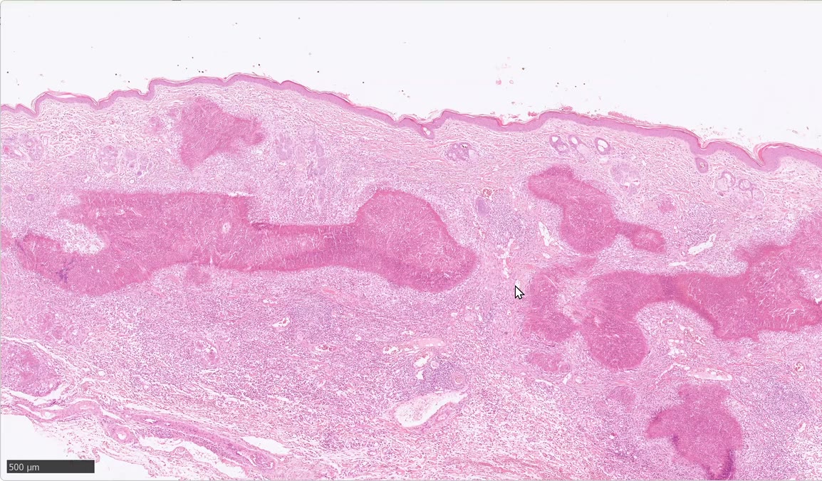
學員描述: 真皮內到處是大結節狀構造=脫顆粒的嗜伊紅球+膠原纖維=flame figures;周圍嚴重瀰漫性嗜伊紅球為主+巨噬細胞浸潤,淺真皮到深真皮、達 panniculus;深真皮少量紅血球(Monika:切片假影)。學員診斷:eosinophilic granuloma。
Monika 的教學:
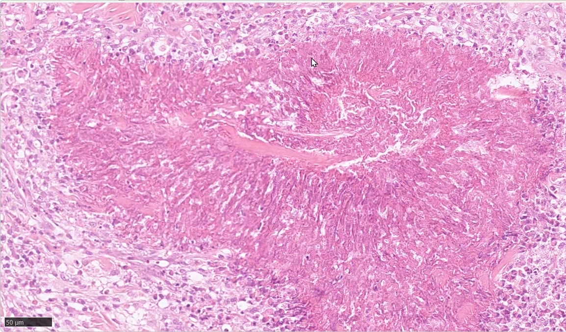
- 寫法:multifocal to coalescing nodular eosinophilic structures = flame figures。是不是 degenerate collagen 「看你信誰」——她認為兩者都有(這麼多嗜伊紅球的蛋白酶,膠原一定會變性)。
- ⭐ Plaque vs granuloma 的判準: flame figure 周圍有沒有 epithelioid macrophages/multinucleated giant cells(或 Langhans cells)圍繞。只有瀰漫性嗜伊紅球 → 還是 eosinophilic plaque;有組織球圍成圈 → 才叫(collagenolytic)eosinophilic granuloma。 本例大部分區域是 plaque,局部(⏱ 15:00)已看到 epithelioid macrophages 與巨細胞圍住 flame figure → 正在轉成 granuloma 的邊界病例。
- 學員問:「plaque 的表皮應該有更多 spongiosis 吧?」→ 不是。 Spongiosis 可出現在任何真皮反應型態(granuloma、plaque、mucinosis…),不能拿來分 plaque 與 granuloma。
- 顏色的說法(Bob Dunstan,C.L. Davis 會議):flame figure 中偏藍=變性的嗜伊紅球(還看得到核殘影);偏紅=變性膠原為主。另有西班牙團隊主張膠原其實正常——她個人不信,但沒研究過。
- 「palisading」一詞:保留給組織球排列;嗜伊紅球在病灶邊緣堆積看起來也像柵欄,那只是病灶「長大」的過程(中心先有病灶、嗜伊紅球一波波來、在邊緣變性)。
- 病灶旁的小淋巴濾泡=對發炎的反應。

🔬 Morph/診斷(依逐字稿補)
- Morph(依學員描述+Monika 修正整理):Superficial to deep dermis, extending to panniculus: multifocal to coalescing nodular eosinophilic structures (flame figures), surrounded by a severe diffuse infiltrate of eosinophils and some macrophages; focally, epithelioid macrophages and multinucleated giant cells surround flame figures; small lymphoid follicle as a reaction to the inflammation.
- 診斷:Feline eosinophilic plaque(eosinophilic granuloma complex),局部已轉成 collagenolytic eosinophilic granuloma 的邊界病例;學員原診斷 eosinophilic granuloma → Monika 認為整體仍應稱 plaque。
🎯 教學重點(依逐字稿補)
- 要用的詞是 flame figures(“what is the term here which we have to use is flame figures”)。
- 叫不叫 granuloma,看的是組織球(epithelioid macrophages/giant cells)有沒有圍住 flame figure,不是看嗜伊紅球多寡。
- 深真皮的少量紅血球=切片假影(biopsy artifact),不必寫成出血。
對照:2025.11.21 ECVD HistoRound Case 8(Sonya:貓 flame figures + mural folliculitis → 想 herpes)|2025.12.13 ECVD HistoRound Case 4(犬 Wells-like,flame figures)|2026.08.22 Board Review Part 1(Trainor 的 feline eosinophilic plaque Key Points)。
❓ Q&A — Elastosis(⏱ 20:59–22:30)
- Solar elastosis 的彈性纖維:非常細、長的絲狀;「藍書」(Gross et al.)稱之為 「Walder’s spaghetti」(Emily Walder 首先描述)——想成義大利麵:細而長(通常不是藍色)。太粗的就不是。
- Monika 找了 20–30 張國外玻片都找不到漂亮的例子;找到會分享。
🎯 本段考點
- 寄生蟲一定要量大小、描述構造(沒尺就拿淋巴球 8–9 µm 當比例)。
- Besnoitia 囊 200–800 µm:壓扁膠原環 → 宿主細胞 → 香蕉形 bradyzoites。
- Eosinophilic plaque vs granuloma = 有沒有組織球/巨細胞圍著 flame figure;spongiosis 不能拿來分。
- Palisading 是組織球的詞,別用在嗜伊紅球。
- Walder’s spaghetti = solar elastosis 的細長彈性纖維。
- 牛:深層叫 hypodermis。
🔑 關鍵字
- Feline eosinophilic plaque(若有)|DermPath
- 系列:2025.11.06 ECVD HistoRound → 2026.02.06 ECVD HistoRound → 本場 → 5/7 Perivascular|ECVD residents 官方錄影庫(dermatology4animals)
❓ 我的問題
- 3 月這場前面 1 小時多(膠原/彈性纖維等細胞外基質的真皮反應型態、「musculopathy」)官方沒錄到——能否向 Monika 要 Dropbox 裡的玻片清單自己看?
- 4 月的 depositions 那場官方頁面沒有——有錄影嗎?
🧠 自我測驗(board 複習用,先想再點開)
Q1. 見 Case 1 第一張圖:牛頭部皮膚,低倍下真皮內大量卵圓形囊狀構造。請寫出完整描述(位置、數量用詞、大小、構造、發炎)與診斷。
答
- 位置:superficial dermis 為主,延伸到 hypodermis。
- 數量:abundant(約 50% 真皮被囊取代)。
- 大小:約 200–800 µm。
- 構造:淡嗜伊紅環(壓扁的纖維結締組織)→ 壓扁的宿主細胞(含胞漿)→ 數百個香蕉形 bradyzoites。
- 發炎:輕微,lymphocytes 為主,另有 eosinophils、histiocytes。
- 診斷:Bovine besnoitiosis。 → 見 Case 1
Q2. 切片上沒有比例尺時,怎麼估計寄生蟲大小?Besnoitia 的囊與 bradyzoite 各約多大?
答
- 用**淋巴球(約 8–9 µm)**當內建比例尺來比較。
- 囊:約 200–800 µm(沒有其他寄生蟲囊這麼大)。
- Bradyzoite:約 5–7 µm,比淋巴球略小。 → 見 Case 1
Q3. 見 Case 1 第二張圖:Besnoitia 囊周圍那圈淡嗜伊紅環是什麼?由外而內依序是哪些構造?
答
- 淡嗜伊紅環=被壓扁的真皮纖維結締組織(學員只說「fibrosis」,Monika 澄清是 compressed fibrous connective tissue)。
- 由外而內:壓扁的結締組織 → 壓扁的宿主細胞與胞漿 → 數百個香蕉形 bradyzoites;其餘都是宿主組織。 → 見 Case 1
Q4. 為什麼牛 Besnoitia 囊數量這麼多,周圍發炎卻相對輕微?
答
- 因為蟲在細胞內(intracellular),而且位在真皮。 → 見 Case 1
Q5. 牛的皮膚切片,深層脂肪/結締組織應該叫什麼?界線在哪?
答
- 叫 hypodermis,不叫 panniculus(性質類似 panniculus)。
- 大約從 anagen 毛球所在處以下就是 hypodermis。 → 見 Case 1
Q6.(選擇題)一張切片約 50% 真皮被寄生蟲囊取代,描述數量時最適合用哪個詞? (A) multiple (B) scattered (C) abundant (D) few
答
- (C) abundant。
- Monika:multiple 給人「很多但零星分布」的印象;一半真皮都被取代就該說 abundant。 → 見 Case 1
Q7. 見 Case 2 第一張圖:貓真皮內大量結節狀嗜伊紅構造。請寫出 morph,並說明這些構造該用什麼術語。
答
- Superficial to deep dermis,延伸到 panniculus:multifocal to coalescing nodular eosinophilic structures(flame figures),周圍有 severe diffuse eosinophilic(及少量 macrophages)浸潤。
- 術語一定要用 flame figures=脫顆粒的嗜伊紅球+膠原纖維。 → 見 Case 2
Q8. Feline eosinophilic plaque 與(collagenolytic)eosinophilic granuloma 在組織上怎麼區分?本例(見 Case 2 第三張圖,⏱ 15:00)屬於哪一種?
答
- 判準:flame figure 周圍有沒有 epithelioid macrophages/multinucleated giant cells(或 Langhans cells)圍繞。
- 只有瀰漫性嗜伊紅球 → eosinophilic plaque;有組織球圍住 → eosinophilic granuloma。
- 本例大部分是 plaque,局部已出現 epithelioid macrophages 與巨細胞圍住 flame figure → 介於 plaque 與 true collagenolytic granuloma 之間的邊界病例;整體 Monika 仍稱 plaque(學員原答 eosinophilic granuloma)。 → 見 Case 2
Q9.(講師糾正的錯誤)學員認為「eosinophilic plaque 的表皮應該有比較多 spongiosis,可以拿來和 granuloma 區分」。這個說法對嗎?
答
- 不對。 Spongiosis 可以出現在任何真皮反應型態(true granuloma、plaque,前面看過的 musculopathy 也有)。
- 所以 spongiosis 不能用來區分 plaque 與 granuloma。 → 見 Case 2
Q10. 見 Case 2 第四張圖:flame figure 有的地方偏藍、有的偏紅,依 Bob Dunstan 的說法各代表什麼?Monika 對「膠原是否變性」的看法是?
答
- 偏藍(還看得到核殘影)=變性的嗜伊紅球;偏紅=以變性膠原纖維為主(Dunstan 在 C.L. Davis 會議所教,Monika 說不確定真假)。
- 有西班牙團隊主張這類病例膠原都正常;Monika 個人不相信——她認為兩者都有:大量嗜伊紅球帶有很多蛋白酶,膠原一定會變性(但她沒有自己研究過)。 → 見 Case 2
Q11. 嗜伊紅球在 flame figure 邊緣排成一圈、看起來像柵欄,可以寫成 palisading 嗎?為什麼會長成這樣?
答
- Palisading 這個詞保留給組織球圍繞病灶的排列。
- 嗜伊紅球邊緣堆積是病灶逐漸長大的過程:先有一個小病灶,嗜伊紅球一波波過來、在那裡變性,看起來才有點像 palisading 或 radiating。 → 見 Case 2
Q12.(選擇題)關於 solar elastosis 的彈性纖維(「Walder’s spaghetti」),下列何者正確? (A) 粗而短、強嗜鹼性 (B) 細而長的絲狀,通常不是藍色 (C) 一定呈藍色團塊 (D) 越粗越典型
答
- (B)。藍書(Gross et al.)稱為 Walder’s spaghetti(Emily Walder 首先描述)——想成義大利麵:細而長,通常不是藍色;太粗的就不是。 → 見 Q&A — Elastosis