2026.02.06 ECVD HistoRound — Group 1「Epidermal reaction patterns」(Monika Welle)
官方標題:Group 1 MW epidermal reaction patterns February。歐洲 2026-02-05(四)=台灣 2/6 凌晨——就是我 oCam 的
2026.02.06_0017/_0027那場(我的側錄只有 4 張截圖+2 句話,_0027那兩句「two nice apoptotic keratinocytes, the nucleus is already breaking apart」正是本片 25:33)。已由官方完整版取代。 🙋 ⏱ 13:41 Monika 問等候室裡的「guest」是誰——「I’m from Taiwan, second-year resident」→ 那是我;她納悶為何我被標成 guest 而不是 resident(可能要請 ECVD 修正名單)。 上個月(1 月)講的是 actinic keratosis(基底/基底上 dysplasia)與 psoriasiform——那場官方頁面播放器是空的。下個月:dermal reaction patterns。 逐字稿:逐字稿/2026.02.06 官方 Group 1 Feb 逐字稿(英文原文).txt;截圖:attachments/2026.02.06_official/(右側 Zoom 欄已裁掉)。
⭐ 本場考點速記(30 秒複習)
- Interface dermatitis(界面性皮膚炎)最好用的線索是 loss of the basal cell layer(基底細胞層消失)+ basal bubbling;盤狀紅斑狼瘡(discoid lupus erythematosus, DLE)的 apoptotic keratinocytes 其實很少、要拚命找——Monika:“it makes it so much easier” → Case 2
- 多形性紅斑(erythema multiforme, EM):apoptotic keratinocytes numerous、transepidermal、伴 satellitosis,在 infundibulum 內層可融合成整段壞死;切片無法區分 EM 與 Stevens-Johnson syndrome/toxic epidermal necrolysis(SJS/TEN)→ Case 3
- Bowenoid in situ carcinoma(BISC,Bowen’s)=full-thickness epidermal dysplasia + abrupt transition + 延伸入 infundibulum;actinic keratosis(AK)=基底/基底上 dysplasia、漸進過渡。“the most important term is really here the dysplasia” → Case 4
- Ulceration=基底膜被破壞;erosion=只有表淺表皮缺損(角質層不能潰瘍);貓糜爛潰瘍+嗜伊紅球浸潤+核內包涵體 → herpes,千萬別誤診成 eosinophilic plaque → Case 5
- 燙傷=full-thickness epidermal coagulation necrosis(全層凝固性壞死):細胞細節全無、核淡或消失、角質層相對保留——不是假影、不是水疱病,也無法判斷原本表皮厚度 → Case 1
- 用詞陷阱:Sarcoptes 在角質層內的是 cavity/burrow,不是 cyst(要寫蟎大小);Pautrier’s microabscess 是誤稱(沒有壁、不是嗜中性球)=表皮內腫瘤性淋巴球團塊 → epitheliotropic lymphoma → Case 6、7
- 已複習
📋 本場案例一覽(7 張玻片=表皮反應型態總複習)
| # | 玻片 | ⏱ | 物種 | 反應型態/診斷 | 一句話重點 |
|---|---|---|---|---|---|
| 1 | 1747 | 03:09–15:40 | 犬 | Full-thickness epidermal coagulation necrosis(燙傷) | 表皮「薄到剩一層、沒細胞細節、整片剝離」=凝固性壞死,不是萎縮也不是假影 |
| 2 | B622 | 16:50–37:40 | 犬 | Interface/cytotoxic dermatitis → Discoid lupus | ⭐ loss of basal cell layer + basal bubbling 比找 apoptotic cell 好用 |
| 3 | B610 | 37:41–53:00 | 犬 | Erythema multiforme + 表面大量 Malassezia | transepidermal apoptosis、satellitosis、spongiosis;花生形酵母菌 |
| 4 | B194 | 54:00–65:00 | 貓 | Bowen’s(BISC):full-thickness epidermal dysplasia | 與 actinic keratosis 的差別:全層+abrupt transition+延伸入 infundibulum |
| 5 | B1340 | 66:04–78:00 | 貓 | Herpesvirus dermatitis | erosion vs ulceration 的定義;核內包涵體很難找但很多 |
| 6 | B1898 | 78:00–86:40 | 犬(耳) | Sarcoptes | 角質層內蟎「cavity/burrow」+旁邊的嗜伊紅性微膿疱 |
| 7 | (最後一張) | 86:41–94:05 | — | Epitheliotropic lymphoma:Pautrier’s microabscesses | 名稱是誤稱(沒有壁、不是嗜中性球)——是腫瘤性淋巴球聚集 |
🧫 1. 燙傷:full-thickness epidermal coagulation necrosis(1747,⏱ 03:09–15:40)
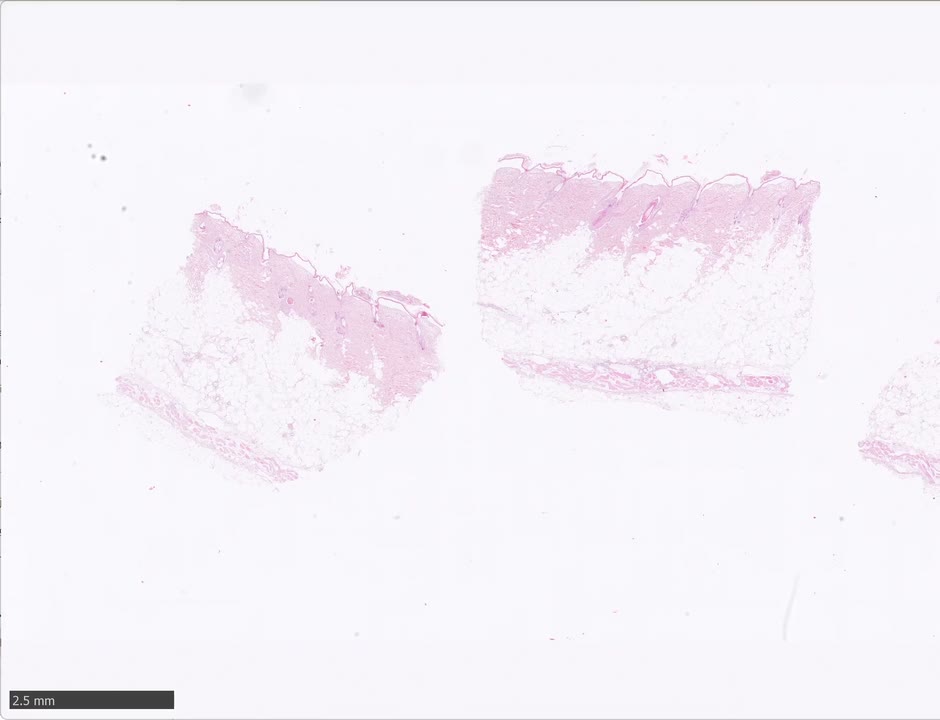
學員(Vanessa)描述: 犬背部有毛皮膚(arrector pili 多);中至局部重度 basket-weave orthokeratotic hyperkeratosis;表皮嚴重變薄、約一層厚、延伸進毛囊口;表皮與真皮分離、只在毛囊口處還黏著 → 懷疑假影。
Monika 的修正:
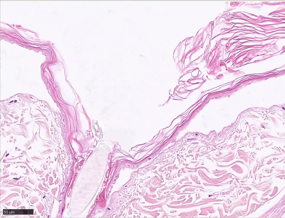
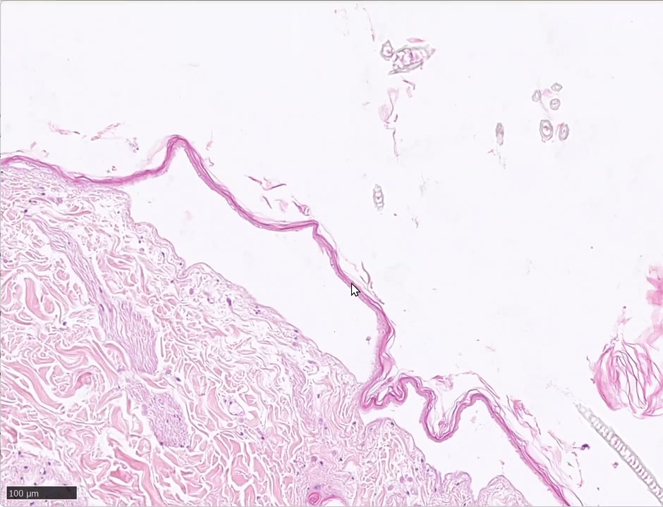
- ⚠️ 表皮這麼薄「有原因」,不要寫 one cell thick;仔細看:看不到任何細胞細節、核極淡或消失(只剩幾個核的殘影)。
- 不是假影——想不出任何假影會長這樣。不是水疱病:subepidermal blistering 時表皮仍完整、細胞細節仍在。
- ⭐ 這是 coagulation necrosis(凝固性壞死):細胞膜被熱破壞、細胞液流失 → 細胞縮成扁片、細節全無 → 所以壞死後無法判斷原本表皮厚不厚。(想想自己燙傷後起水疱、滲液。)
- Stratum corneum 較不受影響(本來就是死細胞,不易「燒壞」)。嚴重時 infundibulum 上皮、甚至真皮也壞死;本例真皮沒有。血管也可見凝固性壞死。
- 來源: 這隻狗被熱水潑到背部。臨床上最常見的燙傷是手術時的電熱毯——溫度太高是一種原因,但更常是狗被移動、上真皮位移、血管受壓 → 局部循環不良 → 很容易燙傷。
- 較舊的燙傷會看到發炎細胞往表面移動修復;本例是早期。
Morph/診斷(依逐字稿補): full-thickness epidermal coagulation necrosis, extending into the follicular infundibula; no cellular details, nuclei very pale or absent; epidermis detached from the dermis(hemidesmosomal anchoring complex 被破壞); coagulation necrosis of blood vessels; stratum corneum relatively spared; dermis not necrotic in this case → Thermal burn(熱水燙傷),early lesion。
🧫 2. Interface/cytotoxic dermatitis → Discoid lupus(B622,⏱ 16:50–37:40)
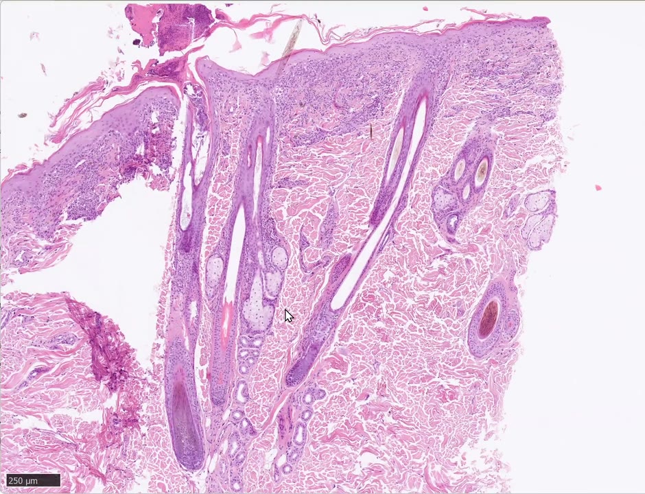
學員(Annika)描述: 犬有毛皮膚;basket-weave(Monika:basket-weave to laminar)orthokeratotic hyperkeratosis;「多層痂」含 keratin 碎屑與嗜中性球(Monika:只有中間夾著一層完整角質才叫 multi-layered,這裡是一個大的 serocellular crust);一處疑似 subcorneal 膿疱/水疱(Monika:只是掀起來的角質,不算;另一處可算 intraepidermal vesicle);真皮表皮交界的發炎、基底層被淋巴球侵入 → cytotoxic interface;基底層 intercellular edema/hydropic change;延伸入 infundibulum(表淺部,仍看得到 keratohyalin);少數 apoptotic keratinocytes(很紅、核碎裂);pigmentary incontinence;大的 subbasal/intrabasal cleft。
Monika 的教學:
- 細胞進入表皮或毛囊上皮的專有名詞=exocytosis(不是 epidermotropism);要說在哪一層:這裡是 basal and suprabasal exocytosis of lymphocytes(不是 transepidermal)。
- 基底細胞成排的 hydropic degeneration=basal(suprabasal)bubbling。
- ⭐⭐ 她「很晚才學到、但讓事情變得容易很多」的訣竅:找 loss of the basal cell layer。 正常基底層是「核、核、核」排成一列的立方細胞;cytotoxic attack 後 apoptotic cells 很快死掉、消失 → 剩下的是沒有基底細胞的一段(或被壓扁)。教科書都寫「找基底層 apoptotic cells」,但 lupus 的 apoptotic cells 其實很少、要拚命找;正常表皮偶爾也有 1–2 顆。看到 1–2 顆 apoptotic cell + 基底層消失 → 可以確定有 cytotoxic attack = interface。
- Cleft 的成因:基底細胞帶著 hemidesmosome 一起消失 → 真皮表皮分離。
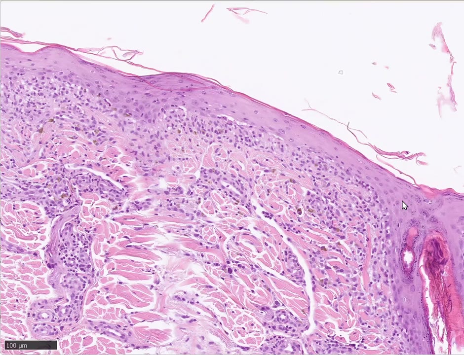
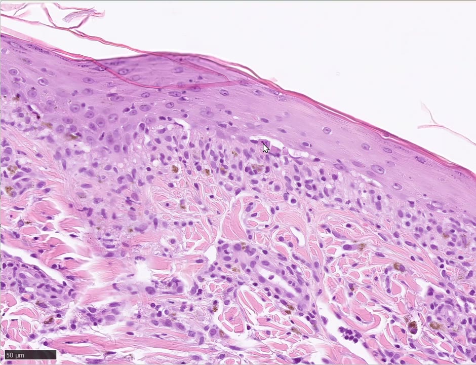
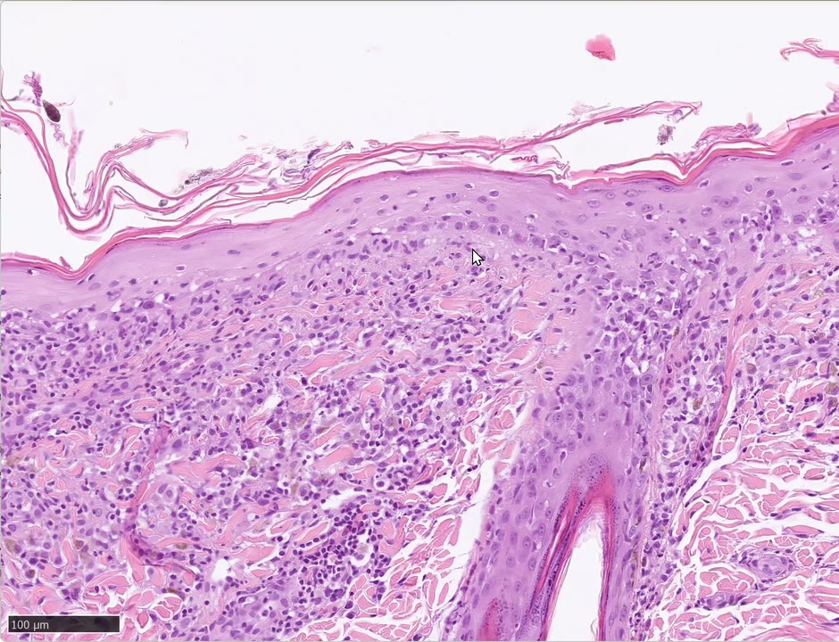
本例特徵總結: interface/cytotoxic dermatitis with basal bubbling, apoptotic keratinocytes, basal ± suprabasal lymphocytic exocytosis, loss of basal cell layer, intraepidermal cleft formation, laminar orthokeratotic hyperkeratosis, serocellular crust;同樣變化在 infundibulum。
Q:這是哪一型 lupus?(⏱ 33:31)
普通的 discoid lupus。DLE 也可有小水疱(vesicular lupus 更明顯)。Monika 的立場:光靠組織病理不能分 lupus 的亞型,一定要臨床資訊(品種、部位、病灶年齡)。她看過的 vesicular lupus(5–6 例):小水疱多、角質細胞常呈方形而非圓形。與 mucocutaneous pyoderma 也難分——那裡也可見 apoptotic cells 與淋巴球(不只漿細胞)。「Lupus 在組織病理上是個難病。」
🧫 3. Erythema multiforme + Malassezia(B610,⏱ 37:41–53:00)
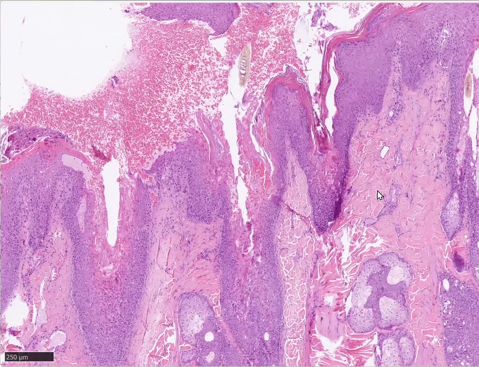
Monika:「描述上這張跟 B1340 是最難的兩張。」
學員(Abigail)描述 → Monika 修正:
- 犬有毛皮膚;compact orthokeratotic hyperkeratosis(不確定 laminar 還是 compact 就寫 laminar to compact——切片各處不同)。
- 表皮上方的「裂縫」是假影(所以有大量游離紅血球);但表面那些「發炎細胞」——不是!是花生形的 Malassezia(沒有胞漿、依切面呈花生/圓/長橢圓);PAS 會亮起來。
- 「anastomosing rete ridges、psoriasiform」→ Monika:看不到 anastomosing,這是 severe irregular hyperplasia(上個月才看過 psoriasiform,別混)。
- 角質細胞之間的淡色細線=spongiosis(嚴重、基底與基底上更明顯);常伴 hydropic degeneration。
- ⭐ apoptotic cells 不是 occasional,是 numerous,而且transepidermal(各層都有),在 infundibulum 內層甚至融合成 10 顆以上相鄰的凋亡細胞、局部整段壞死。
- 圍繞 apoptotic cell 的淋巴球=satellitosis。
- Exocytosis:確定是淋巴球就寫 lymphocytes,不確定寫 leukocytes(可能混有嗜中性球);Malassezia dermatitis 的標誌之一也是淋巴球 exocytosis。
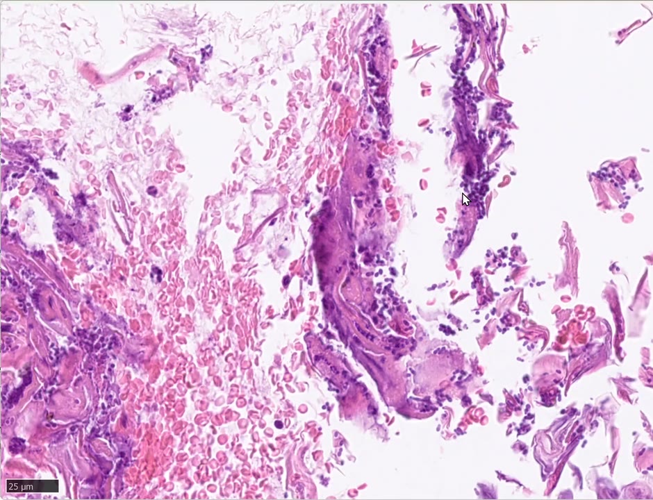
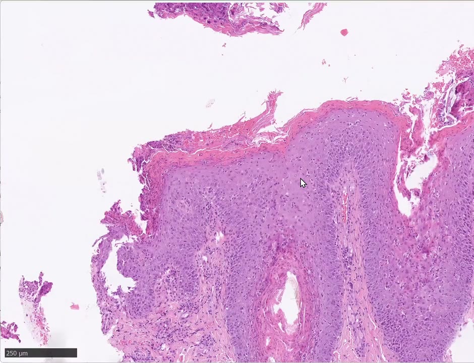
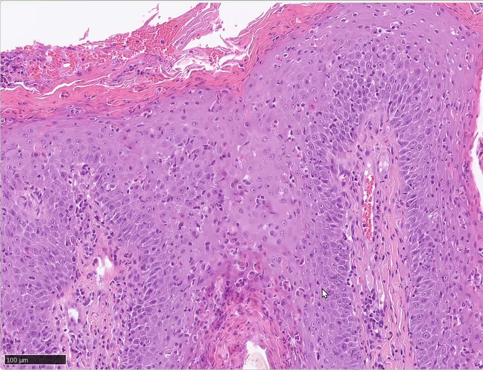
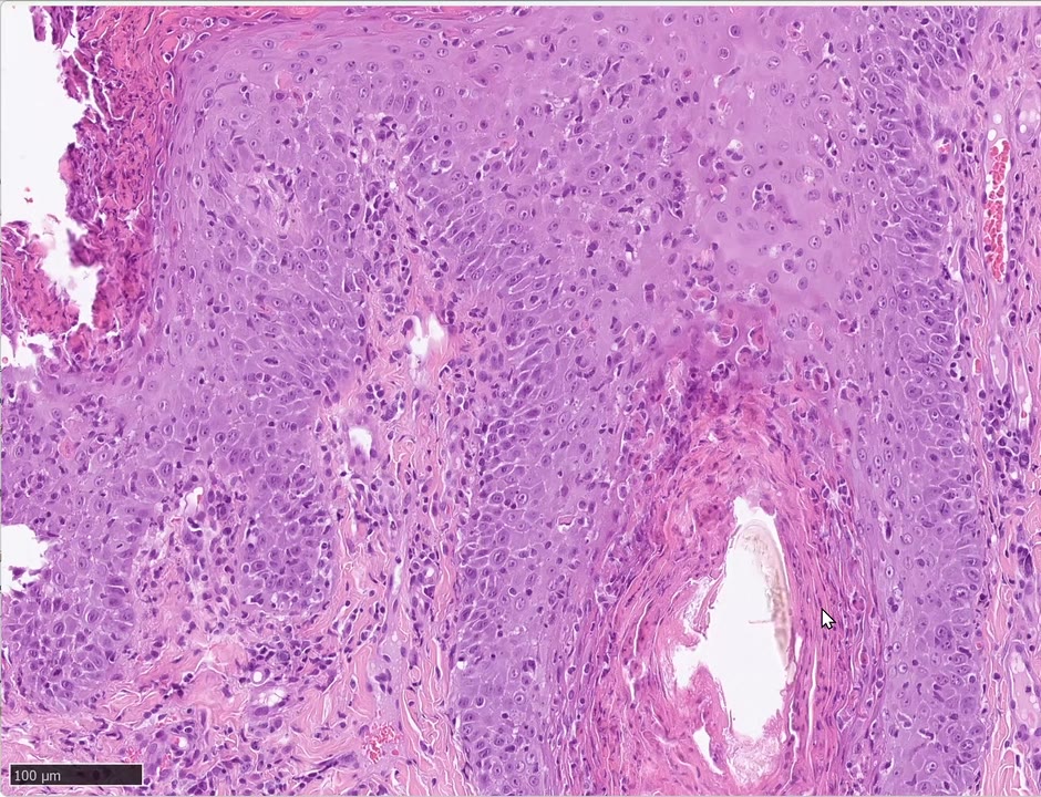
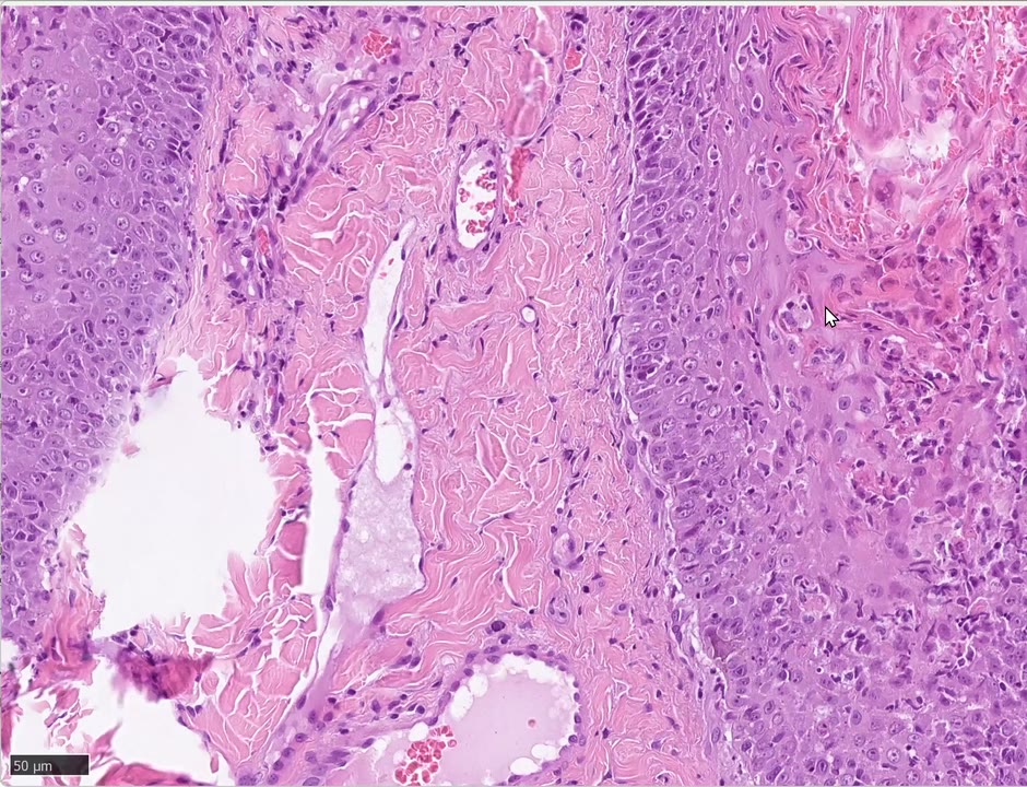
Morph(依逐字稿補,Monika 的總結): laminar to compact orthokeratotic hyperkeratosis; severe irregular epidermal hyperplasia; severe spongiosis(basal and suprabasal 更明顯), hydropic degeneration of keratinocytes; numerous transepidermal and transepithelial(follicular infundibulum, innermost layers)apoptotic keratinocytes, coalescing with focal epidermal necrosis; satellitosis of lymphocytes; exocytosis of lymphocytes(不確定時寫 leukocytes); numerous yeasts(Malassezia)on the surface。“a typical cytotoxic epidermal dermatitis like erythema multiforme”
診斷:Erythema multiforme(重度)。⚠️ 切片不能分 EM 與 SJS/TEN——要看臨床範圍與黏膜是否受累。
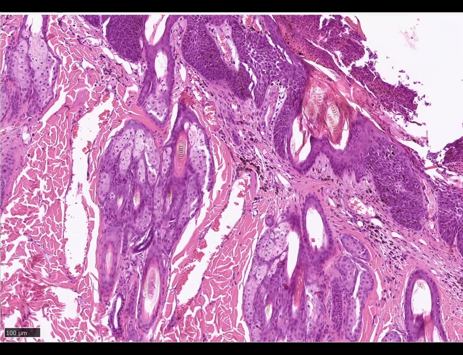
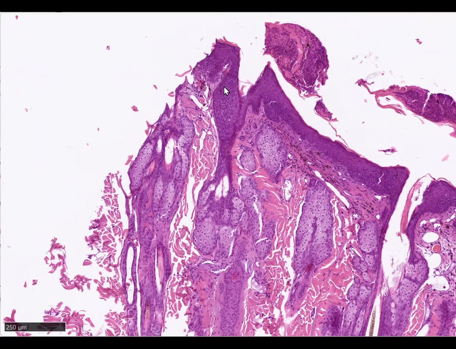
🧫 4. Bowen’s disease(BISC):full-thickness epidermal dysplasia(B194,⏱ 54:00–65:00)
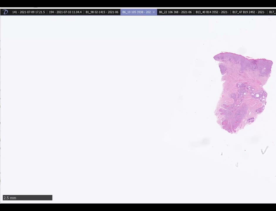
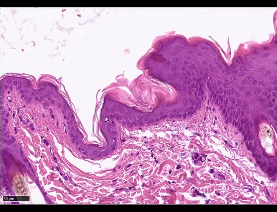
學員描述: 貓有毛皮膚;左側正常區(學員又寫「mild hyperkeratosis」→ Monika:別動不動就寫 hyperkeratosis,貓梳毛多寡差很大,這是正常;表皮 1–2 層也正常);右側局部廣泛的不規則表皮增生、spongiosis、大量黑色素(基底層為主)、出血、serocellular crust(Monika:寫 crust 就好,都是死細胞)。
Monika 的教學:
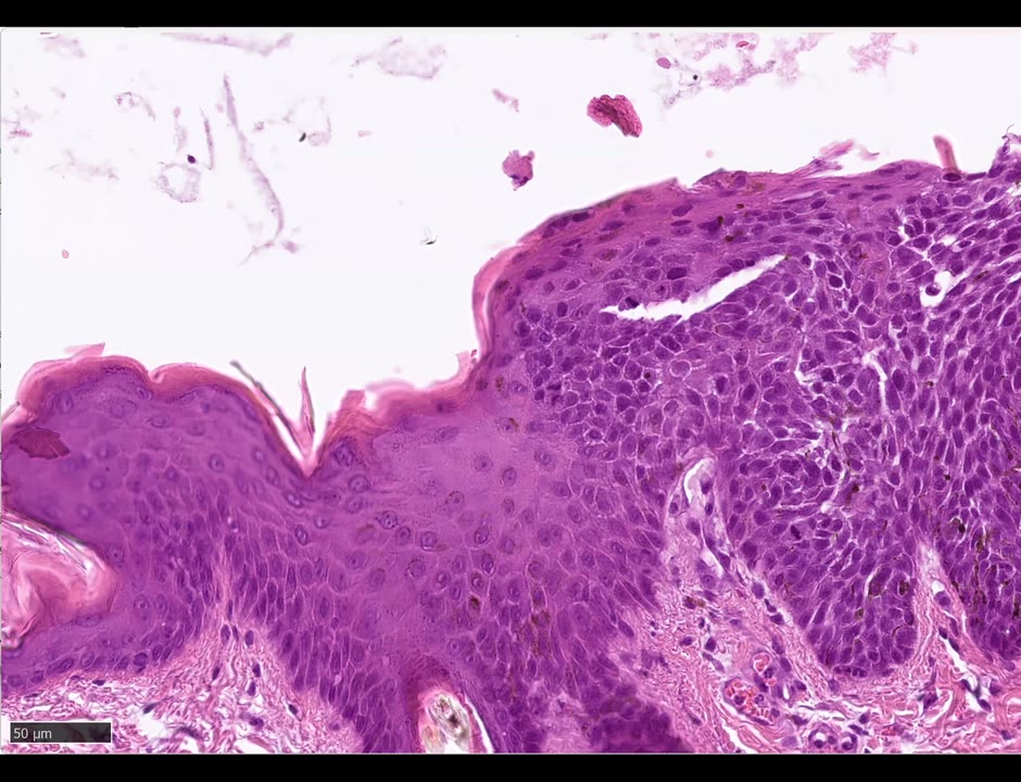
- 病變區表皮比正常區顏色深 → 因為核密度高;而且失去分層(loss of layering):從基底到表面細胞形狀都一樣(略橢圓核)。
- Mitotic figures 在基底上層也有、melanocytes 各層都有、N/C ratio 升高 → epidermal dysplasia。
- ⭐ 與上個月 actinic keratosis 的兩個差別: ① AK 是基底/基底上 dysplasia,BISC 是 full-thickness;② AK 漸進過渡到正常,BISC 是 abrupt transition(本片一側切掉了,另一側應該又突然變正常)。
- 角質細胞朝同一方向「像被風吹的樹/像魚群」=wind-blown appearance。
- 常延伸進 follicular infundibulum(病毒可以進毛囊,陽光只照表面 → AK 較少)。
- 描述模板: 「(multi)focally extensive, full-thickness epidermal dysplasia with abrupt transition to normal epidermis; increased N:C ratio, increased mitotic figures, increased melanocytes and hyperpigmentation, wind-blown streaming, extension into follicular infundibular epithelium」——最重要的字是 dysplasia。
- 病因:papillomavirus → 所以是局部/多發。
對照:2025.11.21 ECVD HistoRound Case 6(Sonya 講的 BISC:infundibulum「懷孕毛囊」+windblown)與 2026.08.06 ECVD HistoRound。
🧫 5. 貓 herpesvirus dermatitis(B1340,⏱ 66:04–78:00)
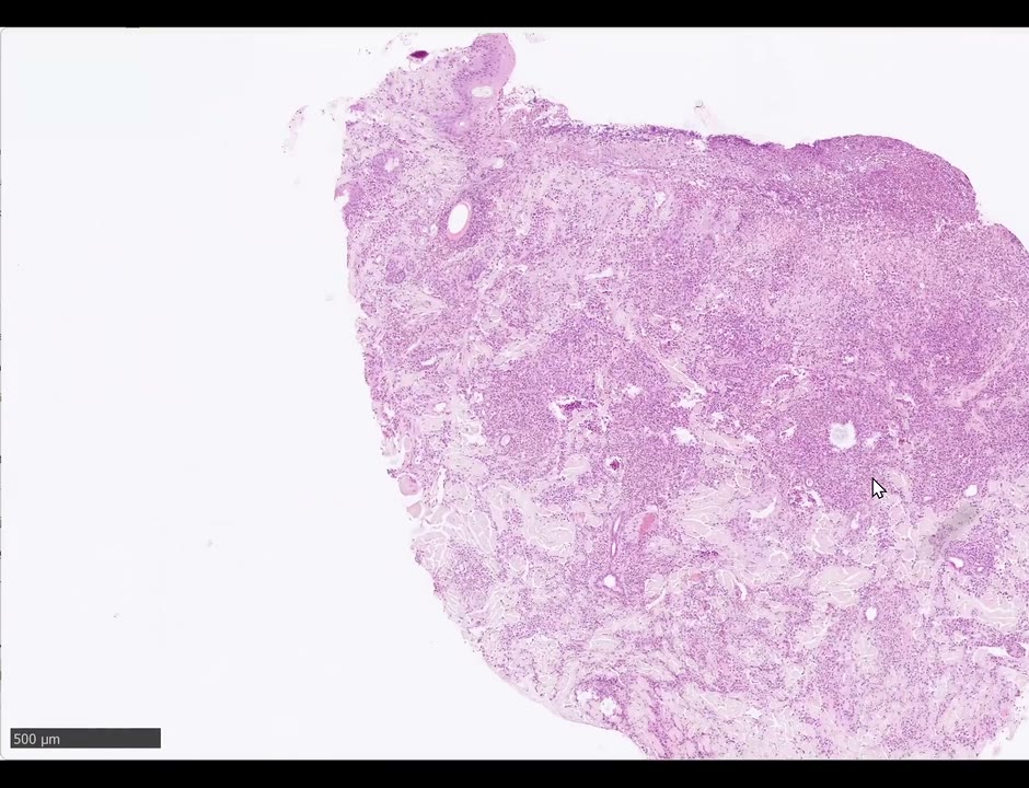
學員(Adrien): 猜狗 → Monika:是貓(「有 20 年經驗看到這種上皮 rafts 就知道是貓」)。中度不規則表皮增生(學員想寫 pseudocarcinomatous → Monika:不到,寫 severe irregular hyperplasia);上層表皮嗜伊紅球為主+少數嗜中性球浸潤;「角質層潰瘍」→ Monika:角質層不能潰瘍。
Monika 放這張的目的:
- ⭐ Ulceration vs erosion:ulceration=基底膜被破壞(基底層+基底膜都沒了、看到裂到真皮);erosion=只有表淺表皮缺損。(對照 Sonya 的講法:erosion 可深到只剩基底層——見 2025.11.21 ECVD HistoRound Case 8。)
- 其他可用術語:spongiosis、hydropic degeneration、exocytosis(貫穿表皮往上到被破壞的表面)。
- ⭐ Intranuclear inclusion bodies:佔滿整個核、核仁消失、染色質被擠到核膜邊緣、核內填滿 amphophilic 物質;「很難看到、要拚命找,第一年不期待你們看到」;在潰瘍邊緣與毛囊最多。
- 哪個病毒造成糜爛潰瘍+嗜伊紅浸潤+核內包涵體 → herpes。 ⚠️ 千萬別誤診成 eosinophilic plaque(治療完全相反)。Bern 第一例(多年前、已發表)就是先被當成 eosinophilic plaque。貓因搔癢也可造成戲劇性糜爛潰瘍,要靠這些小特徵分辨。
Q:包涵體位置速記(⏱ 74:34) —— 核內:herpes、papilloma(也有 pseudo-inclusions,核+胞漿)、parvo、distemper;胞漿內:pox/parapox(巨大、不規則、亮紅、可比細胞還大;pox 潰瘍與真皮壞死很深,包涵體要在深潰瘍旁邊找;浸潤以嗜中性球為主)。
Morph/診斷(依逐字稿補): feline haired skin; severe irregular epidermal hyperplasia(not pseudocarcinomatous); multifocal erosion and ulceration; exocytosis of inflammatory cells(mainly eosinophils, fewer neutrophils)throughout the epidermis toward the destroyed surface; spongiosis and hydropic degeneration; intranuclear amphophilic inclusion bodies in keratinocytes, most numerous at ulcer margins and in hair follicles → Feline herpesvirus dermatitis。
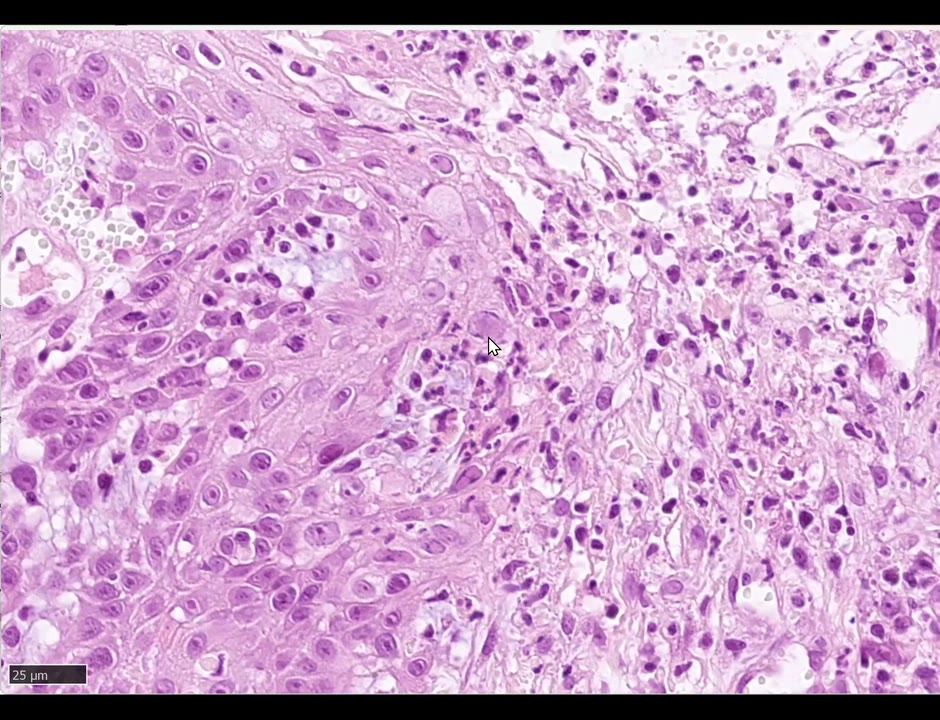
🧫 6. Sarcoptes(B1898,⏱ 78:00–86:40)
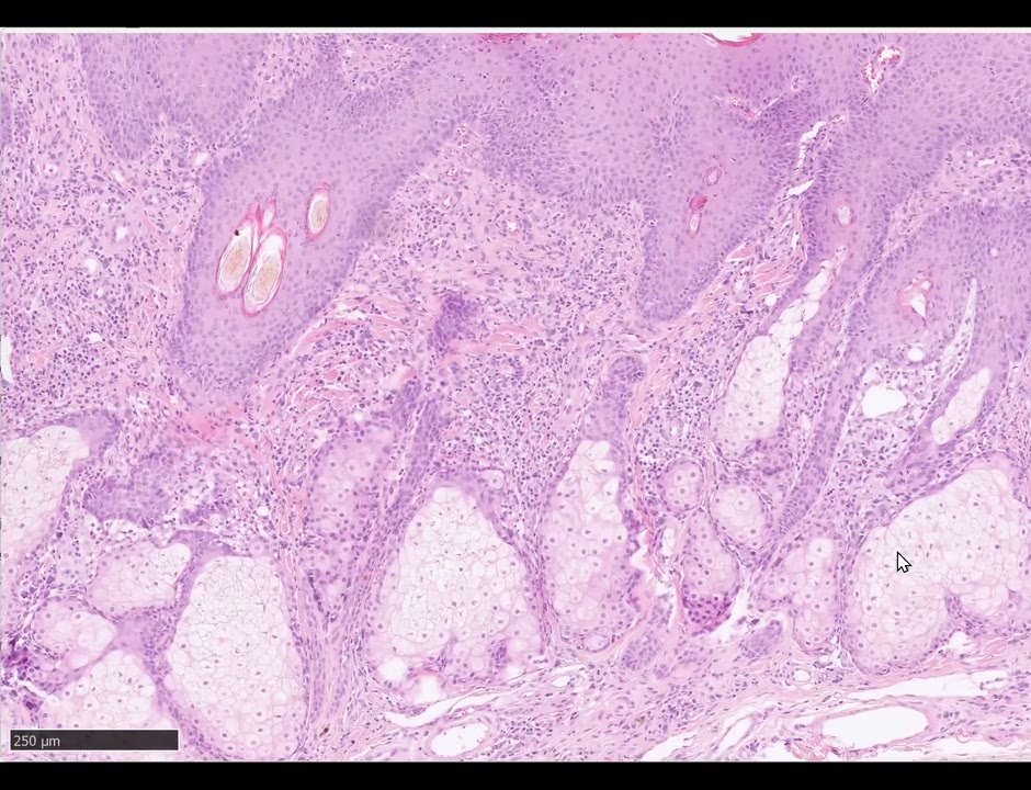
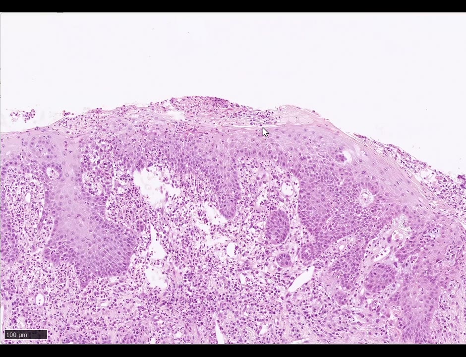
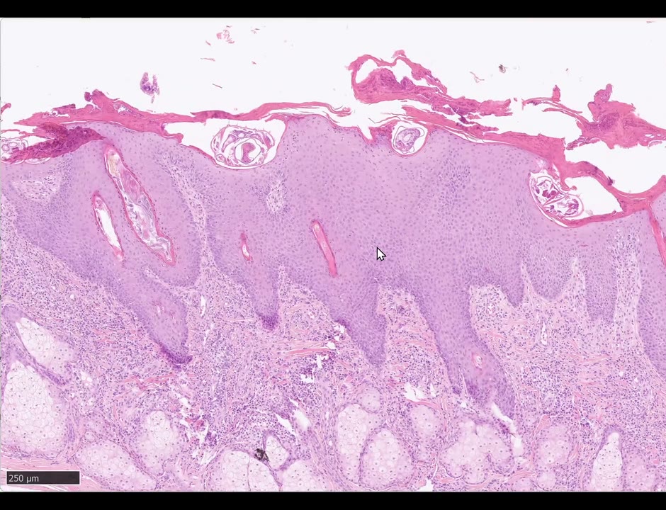
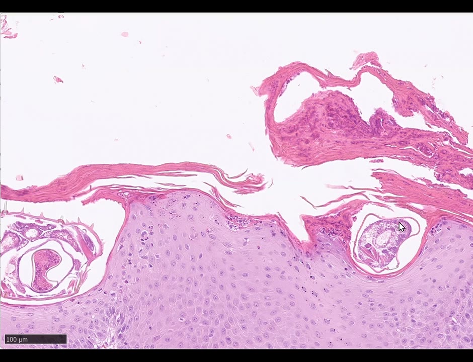
學員(Adrien)描述: 犬耳部有毛皮膚、compound follicles;serocellular crusts;parakeratotic compact hyperkeratosis;角質層內的寄生蟲「囊」、外層有棘刺、150–300 µm;intracorneal 嗜伊紅性膿疱;表皮 pseudocarcinomatous 增生。
Monika 的修正:
- 不是 cyst(cyst 要有壁)——叫 cavity 或 burrow(隧道);看到的是蟎的 cuticle 與棘刺;要寫大小。
- 角質:有些區完全 orthokeratotic → 寫 laminar to compact severe orthokeratotic hyperkeratosis, multifocally parakeratotic。
- 蟎的隧道常被 serocellular crust 覆蓋;蟎旁邊有嗜伊紅球為主的 micropustules + 嗜伊紅球 exocytosis——這些一定要描述。
- Pseudocarcinomatous:學員今天很愛用這詞;Monika 認為多數區域只是 severe irregular hyperplasia(她為 pattern 課找好的 pseudocarcinomatous 例子都很難找)。
- 描述順序建議: 表皮嚴重不規則增生 → 覆蓋的角化 → 角質層內含 X µm 蟎的多個 cavities → 蟎旁的嗜伊紅性微膿疱 → exocytosis。
- 診斷:Sarcoptes。⚠️ Sarcoptes 理論上只在角質層,但她看過侵入活表皮的病例;罕見情況嗜中性球多於嗜伊紅球——別被嚇到。
🧫 7. Pautrier’s microabscess=epitheliotropic lymphoma(⏱ 86:41–94:05)
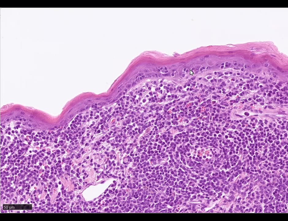
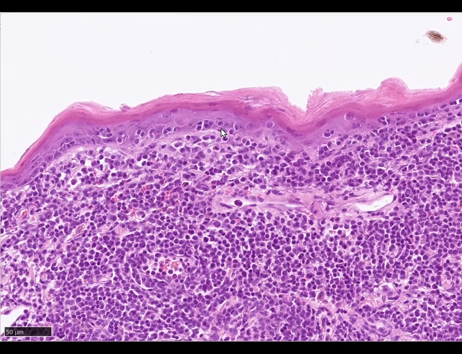
只為了一個名詞:學員描述表皮內淋巴球小聚集+下方大量淋巴球浸潤 → Pautrier’s microabscesses ✅。
- 與 exocytosis(散在穿越)和 satellitosis(圍著 apoptotic cell)不同:這是純淋巴球的團塊,中間沒有 apoptotic cell。
- 是腫瘤性 T 細胞 → epitheliotropic lymphoma(另有專場)。
- ⭐ 「microabscess」是徹底的誤稱:abscess=有壁的腔、內含嗜中性球;這裡兩者皆非。更好的寫法:multifocal intraepidermal aggregates of neoplastic lymphocytes——但書上就叫 Pautrier’s microabscess。
Q:satellitosis 只能用在淋巴球嗎? —— 多半是,但 satellite cell 只是「圍著什麼的細胞」,要指明細胞種類(例:toxic shock syndrome 的 neutrophilic satellitosis)。
Morph/診斷(依逐字稿補,只看表皮): mild compact, multifocally parakeratotic hyperkeratosis(角化型態混合); marked transepidermal exocytosis of lymphocytes; multifocal intraepidermal aggregates of neoplastic lymphocytes(Pautrier’s microabscesses)without central apoptotic cells; heavy lymphocytic infiltrate in the underlying dermis; 本切片其他區有 multifocal ulceration → Epitheliotropic lymphoma(腫瘤性 T 細胞)。
🎯 本場考點速記(表皮反應型態術語總表)
| 術語 | 定義/怎麼用 | 本場例子 |
|---|---|---|
| Coagulation necrosis(full-thickness) | 細胞液流失、細節全無、整片剝離;角質層相對保留 | 燙傷 |
| Exocytosis | 白血球進入表皮/毛囊上皮;要說哪一層(basal/suprabasal/transepidermal) | DLE(basal)、EM(transepidermal) |
| Basal/suprabasal bubbling | 成排的基底細胞 hydropic degeneration | DLE |
| ⭐ Loss of basal cell layer | interface 最好用的線索(apoptotic cell 常很少) | DLE |
| Apoptotic keratinocytes + satellitosis | EM 為 transepidermal、numerous、可融合 | EM |
| Spongiosis | 角質細胞間淡色細線(細胞間水腫);只用於上皮 | EM |
| Hydropic degeneration | 角質細胞內水腫 | DLE、EM |
| Dysplasia | 失去分層、核大小不一、N/C↑、基底上 mitoses、melanocytes 各層 | BISC(全層、abrupt)vs AK(基底、漸進) |
| Wind-blown | 角質細胞同向傾斜 | BISC |
| Erosion vs ulceration | 基底膜是否被破壞 | herpes |
| Intranuclear inclusion | 佔滿核、核仁消失、染色質貼邊 | herpes(核內);pox 胞漿內巨大 |
| Cavity/burrow(非 cyst) | 角質層內蟎的隧道;要寫蟎大小 | Sarcoptes |
| Eosinophilic micropustule | 蟎旁的小膿疱 | Sarcoptes |
| Pautrier’s microabscess | 腫瘤性淋巴球團塊(誤稱) | epitheliotropic lymphoma |
| Hyperkeratosis 的形容詞 | basket-weave/laminar/compact;不確定寫 laminar to compact;別動不動寫 hyperkeratosis | 全場 |
| Multi-layered crust | 痂之間要夾完整角質層才算 | DLE |
| Pseudocarcinomatous | 別濫用;多數只是 severe irregular hyperplasia | herpes、Sarcoptes |
🔑 關鍵字
- Parakeratosis | Stratum corneum | 棘層鬆解 | Superficial pyoderma
- 相關:2025.11.06 ECVD HistoRound(同系列第一場)|2025.11.21 ECVD HistoRound(Sonya 的 BISC/herpes)|2026.08.22 Board Review Part 1(Trainor 的 EM/DLE/FHV Key Points)|ECVD residents 官方錄影庫(dermatology4animals)
❓ 我的問題
- 我在 ECVD Zoom 名單裡被標成「guest」——要不要請 ECVD 辦公室把我改成 resident?
- 1 月那場(actinic keratosis、psoriasiform)官方頁面沒有影片——可以請 Monika 補上或提供 Dropbox?(她說有全體共享的 Dropbox 資料夾放所有玻片)
🧠 自我測驗(board 複習用,先想再點開)
Q1. 見 Case 1 第一張圖:犬背部表皮「薄到只剩一層、看不到細胞細節、與真皮分離,只在毛囊口還黏著」。學員寫 “one cell thick… suspected artifact”。Monika 怎麼修正?morph 該怎麼寫?
答
- 不是假影(想不出任何假影會長這樣),也不是 subepidermal blistering(水疱病時表皮仍完整、細胞細節仍在)
- = full-thickness epidermal coagulation necrosis:熱破壞細胞膜 → 細胞液流失 → 細胞縮成扁片、核極淡或消失
- 所以無法判斷原本表皮厚不厚,不要寫 one cell thick
- 角質層相對保留(本來就是死細胞);可延伸入 infundibulum、血管也凝固性壞死;嚴重時真皮也壞死
- 臨床最常見:手術時的電熱毯——多半不是溫度太高,而是狗被移動、上真皮位移、血管受壓 → 循環不良 → 見 Case 1
Q2.(🔴)Interface dermatitis:教科書都說「找基底層 apoptotic cells」,Monika 說她很晚才學到、但讓判讀容易很多的線索是什麼?為什麼有用?
答
- Loss of the basal cell layer(基底細胞層消失或被壓扁)——正常基底層是「核、核、核」排成一列的立方細胞
- Cytotoxic attack → 先有 apoptotic cells → 細胞死掉後隨時間消失 → 只剩沒有基底細胞的一段
- Lupus 的 apoptotic cells 其實很少;正常表皮偶爾也有 1–2 顆
- 1–2 顆 apoptotic cell + 基底層消失 → 可確定有 cytotoxic attack = interface
- 連帶結果:基底細胞帶著 hemidesmosome 一起消失 → cleft formation → 見 Case 2
Q3.(選擇題)下列敘述何者錯誤? (A) 白血球進入表皮或毛囊上皮的術語是 exocytosis,描述時要說明在哪一層 (B) 淋巴球圍繞 apoptotic keratinocyte 稱 satellitosis (C) satellitosis 只能用於淋巴球 (D) 成排基底細胞的 hydropic degeneration 可稱 basal bubbling
答
- (C) 錯:satellite cell 只是「圍著什麼的細胞」,多半是淋巴球,但要指明細胞種類(例:toxic shock syndrome 的 neutrophilic satellitosis)
- (A) 層別:basal/suprabasal/transepidermal;細胞確定是淋巴球寫 lymphocytes,不確定寫 leukocytes → 見 Case 2、3、7
Q4. 這張 DLE 能不能光靠組織病理分出 lupus 的亞型?需要什麼資訊?還容易跟什麼混淆?
答
- Monika 的立場:不能,一定要臨床資訊——品種、部位、病灶年齡、何時何處切片
- DLE 也可見小水疱;vesicular lupus 小水疱更多、發炎細胞較少、角質細胞常呈方形而非圓形(她只看過 5–6 例)
- 與 mucocutaneous pyoderma 也難分:那裡也可見 apoptotic cells 與淋巴球(不只漿細胞)
- “lupus is really… a difficult disease on histopathology” → 見 Case 2
Q5. 見 Case 3 第二張圖:學員把表皮表面一大片小顆粒當成「inflammatory cells」。其實是什麼?怎麼辨認/確認?跟本例的哪個表皮變化有關?
答
- 大量 Malassezia:沒有像顆粒球的胞漿;依切面呈花生形、圓形或長橢圓
- 過碘酸雪夫染色(periodic acid-Schiff, PAS)會亮起來
- Malassezia dermatitis 的標誌之一也是淋巴球 exocytosis
- 旁邊那道裂縫+游離紅血球=切片假影,不用理 → 見 Case 3
Q6.(案例申論)犬有毛皮膚:嚴重不規則表皮增生、角質細胞間淡色細線、各層都有大量凋亡角質細胞且在 infundibulum 內層融合、周圍有淋巴球、表面大量花生形酵母菌。請寫出完整 morph、診斷與診斷上的限制。
答
- Laminar to compact orthokeratotic hyperkeratosis
- Severe irregular epidermal hyperplasia(不是 psoriasiform:看不到 anastomosing rete ridges)
- Severe spongiosis(基底與基底上更明顯)+ hydropic degeneration
- Numerous transepidermal apoptotic keratinocytes,infundibulum 內層融合(10 顆以上相鄰)、局部整段壞死
- Satellitosis;lymphocytic(或 leukocytic)exocytosis;表面大量 Malassezia
- 診斷:Erythema multiforme(重度)
- 限制:切片不能分 EM 與 SJS/TEN——要看臨床範圍與黏膜是否受累 → 見 Case 3
Q7.(概念比較)DLE(Case 2)與 EM(Case 3)同屬 cytotoxic/interface 型態,表皮變化有哪些差別?
答
DLE(Case 2) EM(Case 3) Apoptotic keratinocytes 很少、要拚命找 numerous,可融合 位置 基底層為主 transepidermal,infundibulum 內層最明顯 Exocytosis basal and suprabasal(非 transepidermal) 全層 其他 basal bubbling、loss of basal cell layer、cleft、pigmentary incontinence satellitosis、severe spongiosis、hydropic degeneration → 見 Case 2、3
Q8.(🔴 概念比較)Bowen’s(BISC)與上個月的 actinic keratosis 都是 epidermal dysplasia,Monika 強調哪兩個差別?BISC 為何常延伸進 infundibulum?
答
- ① 範圍:AK=基底/基底上 dysplasia;BISC=full-thickness(沒有任何一層正常)
- ② 邊界:AK=漸進過渡到正常;BISC=abrupt transition
- BISC 由 papillomavirus 引起(所以局部/多發);病毒可進毛囊,陽光只照表面 → AK 較少延伸 infundibulum
- “the most important term is really here the dysplasia” → 見 Case 4
Q9. 見 Case 4:你怎麼判斷病灶區是 dysplasia?「wind-blown appearance」是什麼?
答
- 病灶區顏色較深=核密度高
- Loss of layering:從基底到表面細胞形狀都一樣(略橢圓到長形核)
- 基底上層也有 mitotic figures、melanocytes 各層都有、N:C 比(nuclear-to-cytoplasmic ratio)升高、hyperpigmentation
- Wind-blown:角質細胞朝同一方向傾斜,像被風吹的樹或一群魚(streaming)
- 附帶糾正:貓左側正常區別寫 mild hyperkeratosis(梳毛多寡差很大);表皮 1–2 層也是正常 → 見 Case 4
Q10.(🔴)Ulceration 和 erosion 的定義差在哪?學員寫「角質層潰瘍」錯在哪?
答
- Ulceration=基底膜被破壞(基底層+基底膜都沒了)
- Erosion=只有表淺表皮缺損
- 角質層不能潰瘍
- 這是 Monika 放 B1340 這張玻片的主要目的 → 見 Case 5
Q11.(🔴)貓皮膚:糜爛潰瘍、嗜伊紅球為主的浸潤。要找什麼特徵才不會漏診?最怕誤診成什麼?
答
- Intranuclear inclusion bodies:佔滿整個核、核仁消失、染色質被擠到核膜邊緣、核內填滿 amphophilic 物質
- 最多在潰瘍邊緣與毛囊;很難看、要拚命找(第一年不期待你看到)
- 診斷:feline herpesvirus dermatitis
- ⚠️ 千萬別誤診成 eosinophilic plaque(治療完全相反);Bern 第一例 herpes 當初就被當成 eosinophilic plaque
- 貓因嗜伊紅性皮膚炎搔癢也會造成戲劇性糜爛潰瘍 → 靠包涵體等小特徵區分 → 見 Case 5
Q12.(選擇題)下列病毒的包涵體位置,何者錯誤? (A) Herpesvirus:核內 (B) Papillomavirus:核內,也有 pseudo-inclusions(核+胞漿) (C) Poxvirus:核內,小而規則 (D) Parvovirus、distemper virus:核內
答
- (C) 錯:pox/parapox 是胞漿內,巨大、不規則、亮紅(嗜伊紅),可比細胞還大
- Pox 潰瘍與真皮壞死很深,包涵體要在深潰瘍旁邊找;浸潤以嗜中性球為主 → 見 Case 5
Q13. 見 Case 6 第一張圖:角質層內有帶棘刺的寄生蟲構造。學員寫 “intracorneal parasitic cysts”——應該怎麼寫?完整描述的建議順序?
答
- 不是 cyst(cyst 要有壁)→ cavity 或 burrow;看到的是蟎的 cuticle 與棘刺;要寫大小(本例約 150–300 µm)
- 順序:表皮 severe irregular hyperplasia → laminar to compact severe orthokeratotic hyperkeratosis, multifocally parakeratotic → 角質層內多個含 X µm 蟎的 cavities(常被 serocellular crust 覆蓋)→ 蟎旁嗜伊紅球為主的 micropustules → 嗜伊紅球 exocytosis
- 診斷:Sarcoptes
- 例外:可侵入活表皮;罕見嗜中性球多於嗜伊紅球——“don’t be irritated by that” → 見 Case 6
Q14. Pautrier’s microabscess 和 exocytosis、satellitosis 有什麼不同?為什麼 Monika 說這名稱是 “a complete misnomer”?比較好的寫法是?
答
- Exocytosis=白血球散在穿越表皮;satellitosis=圍著 apoptotic cell;Pautrier’s=純淋巴球的團塊,中間沒有 apoptotic cell
- 這些是腫瘤性 T 細胞 → epitheliotropic lymphoma
- 誤稱:abscess=有壁的腔、內含嗜中性球,這裡兩者皆非
- 較好的寫法:multifocal intraepidermal aggregates of neoplastic lymphocytes(但書上就叫 Pautrier’s microabscess) → 見 Case 7
Q15.(講師糾正的常見錯誤)以下學員用語各錯在哪? (a) 一個大痂寫成 multi-layered crust (b) 嚴重增生就寫 pseudocarcinomatous (c) 看不到 anastomosing 卻寫 psoriasiform (d) 不確定角質型態時硬選一個
答
- (a) 要兩層痂中間夾一層完整角質層才叫 multi-layered;否則寫 large serocellular crust
- (b) 別濫用;多數只是 severe irregular hyperplasia(Monika 為 pattern 課要找好的 pseudocarcinomatous 例子都很難找)
- (c) Psoriasiform 要有 anastomosing rete ridges;沒有就寫 severe irregular hyperplasia
- (d) 切片各處不同,不確定就寫 laminar to compact(或 basket-weave to laminar) → 見 Case 2、3、5、6