2026.09.13 Q&A + Mock Exam — Tumors 補講+模擬考
講者:Dr. Karen Trainor | 片長 1:04:32 | 看錄影(開放到 2026/11/15)
- 已看完並整理(2026-09-15)
版權
課程投影片截圖僅存於本機 vault 供個人複習,不可外流、分享或再散布(Innovative Vet Path 版權聲明)。
這場在做什麼
最後一堂。前 46 分鐘補完 Part 2 沒講到的腫瘤(7 個)→ 5 題腫瘤小考 → 20 題 mock exam(全出自 Part 1 表皮疾病)→ 開放 Q&A(無人提問)。 🎯 一句話:腫瘤只挑「皮膚科醫師會常碰到、看報告要懂」的 7 個;mock exam 全場幾乎全對 → 這些是基本盤,考試不能錯。
🗺 時間軸
| 時間 | 內容 |
|---|---|
| 00:00–01:43 | 開場,跳過 Part 2 投影片找到腫瘤段 |
| 01:43–46:00 | Tumors:epidermal 3 → follicular 1 → nail bed 1 → fibrous 1 → lymphocytic 1 |
| 46:00–49:16 | 腫瘤小考 5 題(QR code 故障,改口頭) |
| 49:16–52:12 | Q&A 開放、課後問卷說明 |
| 52:12–62:16 | Mock exam 20 題 |
| 62:16–64:32 | 收尾、DermPath 101 預告 |
壹、TUMORS(Part 2 延過來的腫瘤)⏱ 01:43–46:00
Trainor 的選題邏輯(本場開頭原話整理)
腫瘤是整個病理專科天天在啃的東西,皮膚科醫師不需要樣樣精通;她只挑**「看到要認得、拿到病理報告要看得懂、職涯中會常下這個診斷」**的少數幾個。 ⚠️ Part 2 結尾她說「還剩 6 個腫瘤」,實際講了 7 個(多了 metastatic pulmonary carcinoma)。
| # | 腫瘤 | 分類 | 一句話記憶 |
|---|---|---|---|
| 1 | Pigmented viral plaque | Epidermal | 巴哥/雪納瑞腹側黑色斑塊,看不到病毒包涵體 |
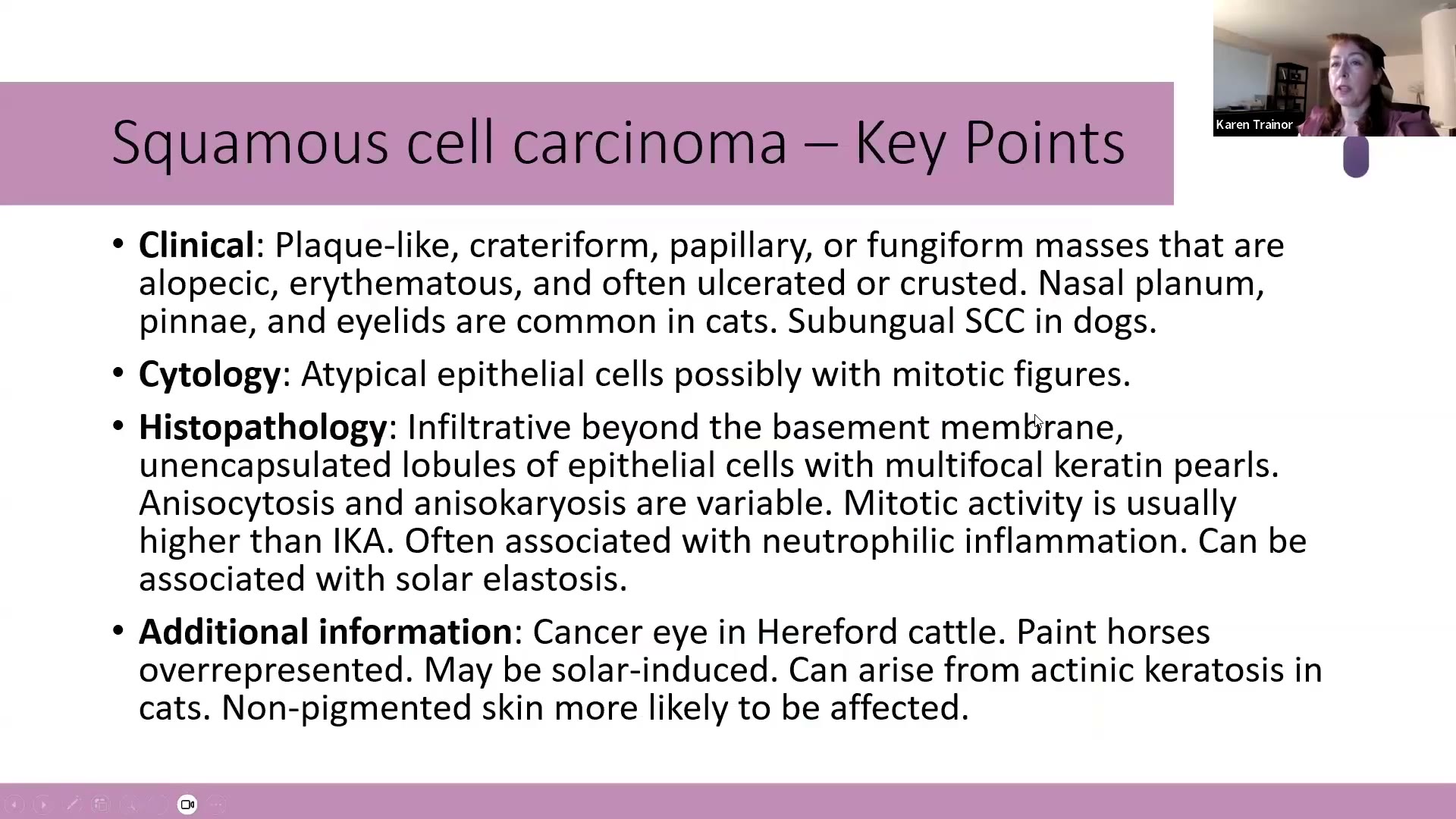
| 2 | Squamous cell carcinoma(SCC,鱗狀上皮細胞癌) | Epidermal | Keratin pearl + solar elastosis;常見病放在罕見物種是命題愛用招 |
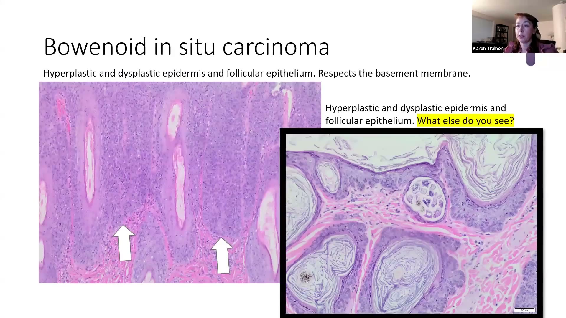
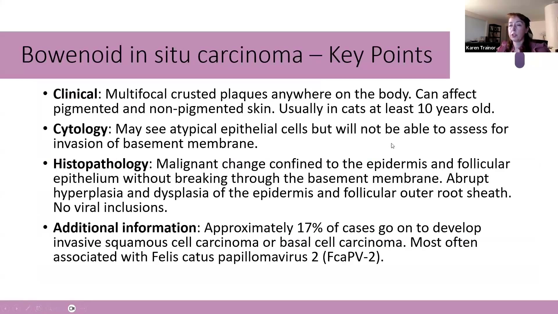
| 3 | Bowenoid in situ carcinoma(BISC,鮑恩樣原位癌) | Epidermal | 老貓多發結痂斑塊,尊重基底膜;FcaPV-2 |
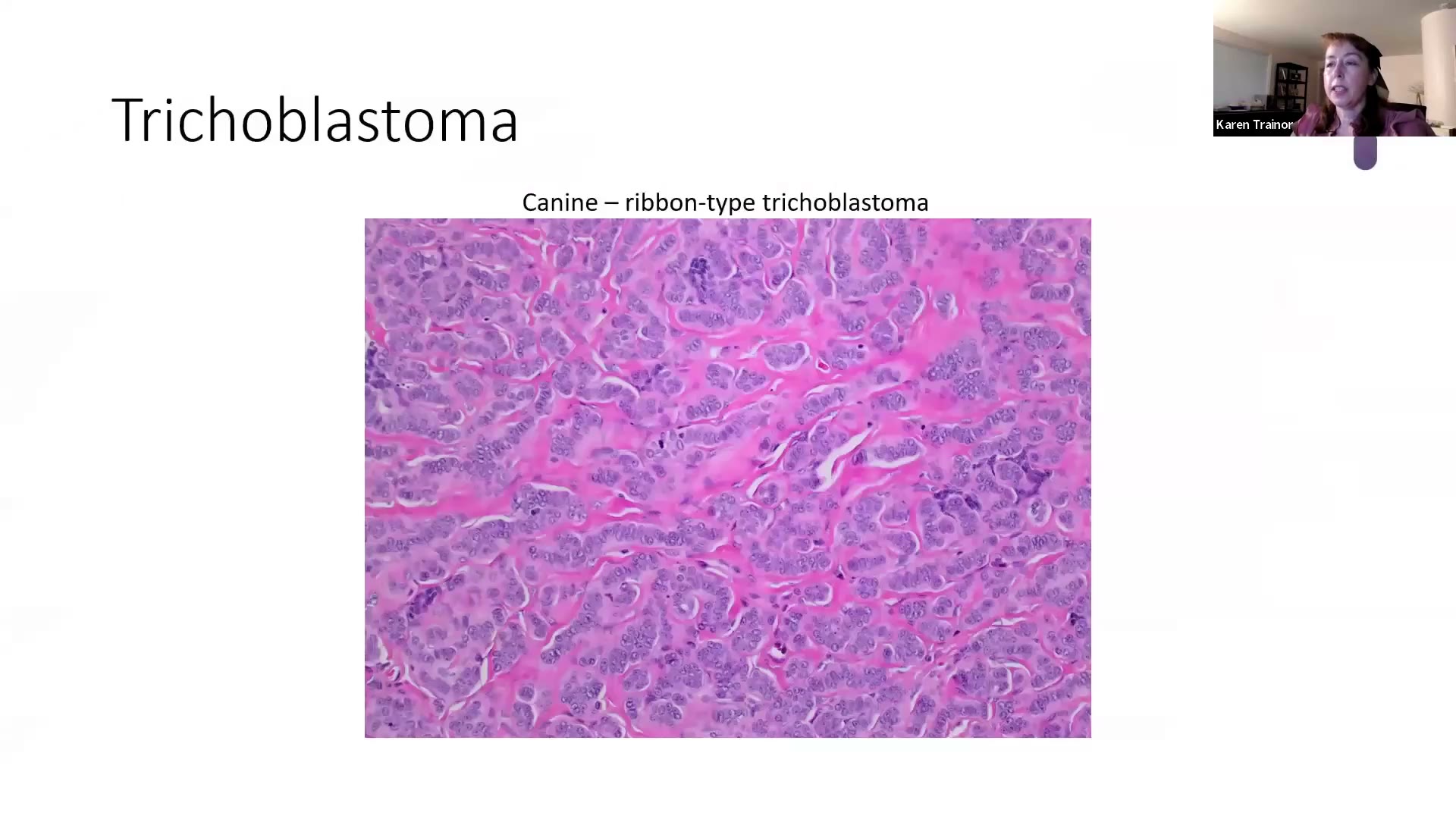
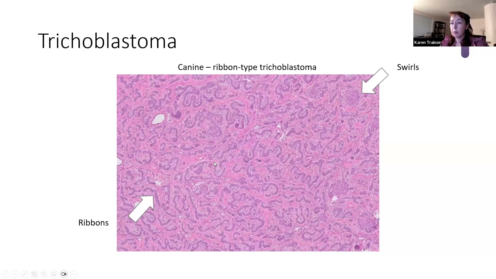
| 4 | Trichoblastoma(毛母細胞瘤) | Follicular | 狗=ribbon type、貓=trabecular type;狗耳根下垂腫塊 |
| 5 | Metastatic pulmonary carcinoma(肺癌轉移至趾,lung-digit syndrome) | Nail bed | 老貓腫趾+ciliated respiratory epithelium |
| 6 | Equine sarcoid(馬類肉瘤) | Fibrous | Picket fence;biphasic → 一定要有完整表皮 |
| 7 | Epitheliotropic lymphoma(親上皮性淋巴瘤) | Lymphocytic | Pautrier microabscesses;鼻鏡—上唇去色素「八字鬍」 |
1. Pigmented viral plaque(色素性病毒斑塊)⏱ 02:57–07:45
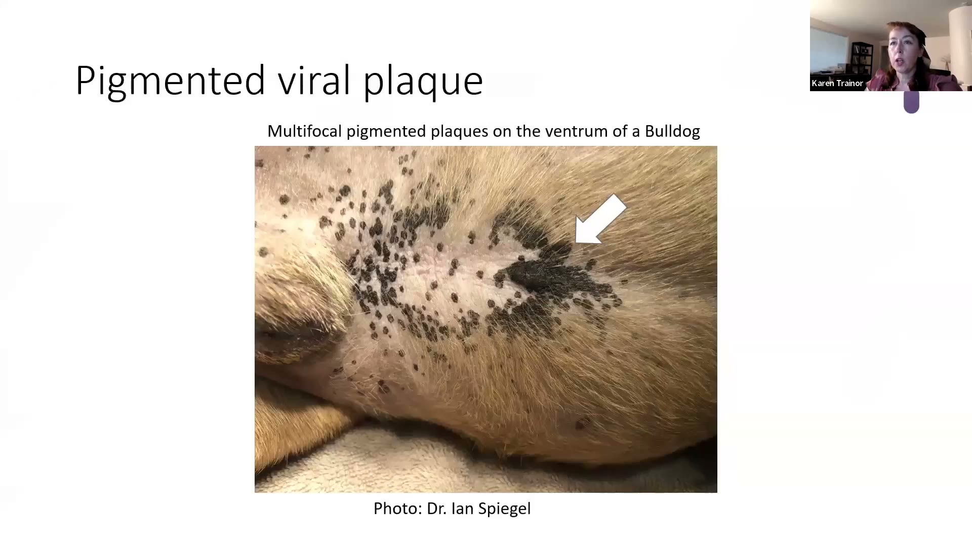
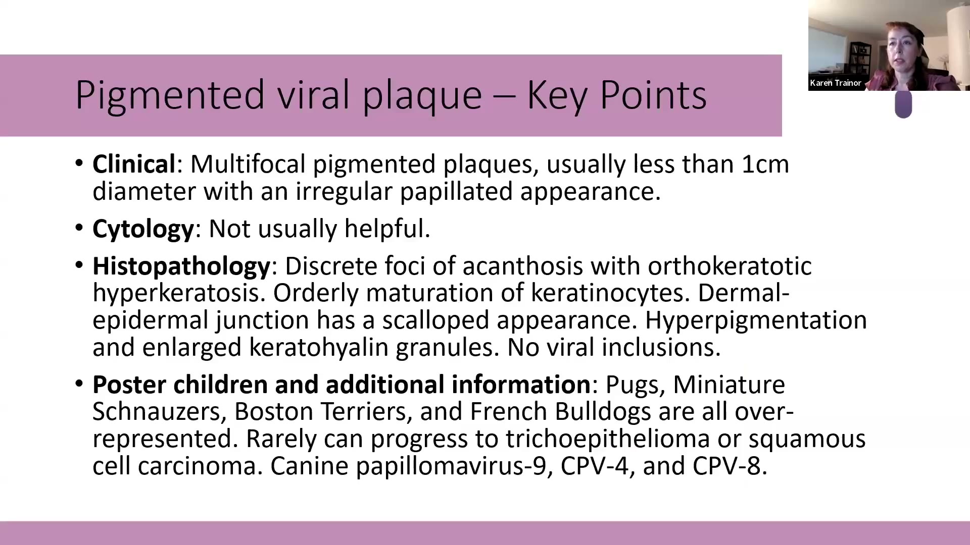
- Clinical:主要在狗;腹側(ventrum)多發性色素斑塊,通常 <1 cm,表面不規則乳突狀,可融合成片。範例是鬥牛犬。
- Cytology:通常沒有幫助(她說若有新文獻請告訴她)。
- Histopathology:
- Papillary(乳突狀)epidermal hyperplasia,外生性(exophytic)突出皮膚表面
- 真皮表皮交界(dermal-epidermal junction)呈扇貝狀(scalloped),不是一條直線
- 表皮突起之間夾著大團 orthokeratotic keratin(她形容是「giant scoops」)
- 色素沉著可從基底層一路到 stratum corneum(角質層裡也有黑色素)
- Granular cell layer 明顯、keratohyalin granules 變大
- 🎯 看不到病毒包涵體(no viral inclusions) —— 不要因為找不到 intranuclear inclusion 就排除病毒斑塊
- Additional:Canine papillomavirus(CPV)9、4、8 最常分離到。好發犬種:pug(巴哥)、miniature schnauzer(迷你雪納瑞)、Boston terrier(波士頓㹴)、French bulldog(法鬥)及其他鬥牛犬。
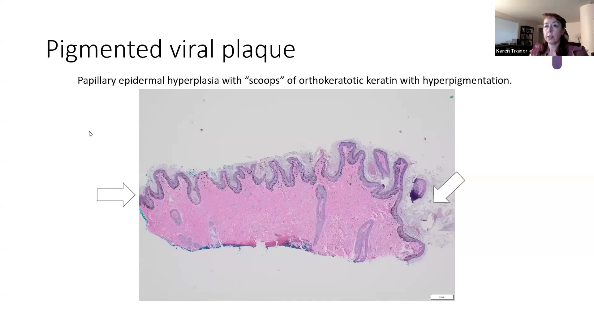
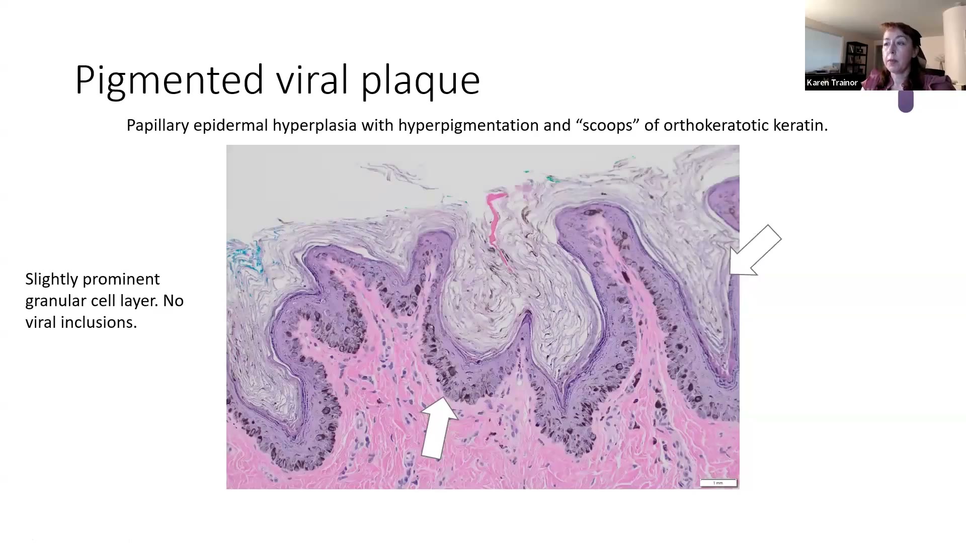
💡 Trainor 的考點與臨床提醒
- 送切片最常見的理由是擔心 melanoma(黑色素瘤)或惡性轉化。絕大多數是良性。
- 什麼時候該切:斑塊不太對勁、融合成大片、或摸起來往下侵入真皮(真皮或皮下出現結節/囊腫樣病灶),不只是浮在表面。
- 罕見會惡性轉化;她看過的轉化病例多半有在用免疫調節藥物。
- 投影片原文:罕見可進展為 trichoepithelioma(毛上皮瘤)或 squamous cell carcinoma。她補充:有時只是良性的 trichoepithelioma,但有時會惡性轉化成較具侵襲性的 SCC。
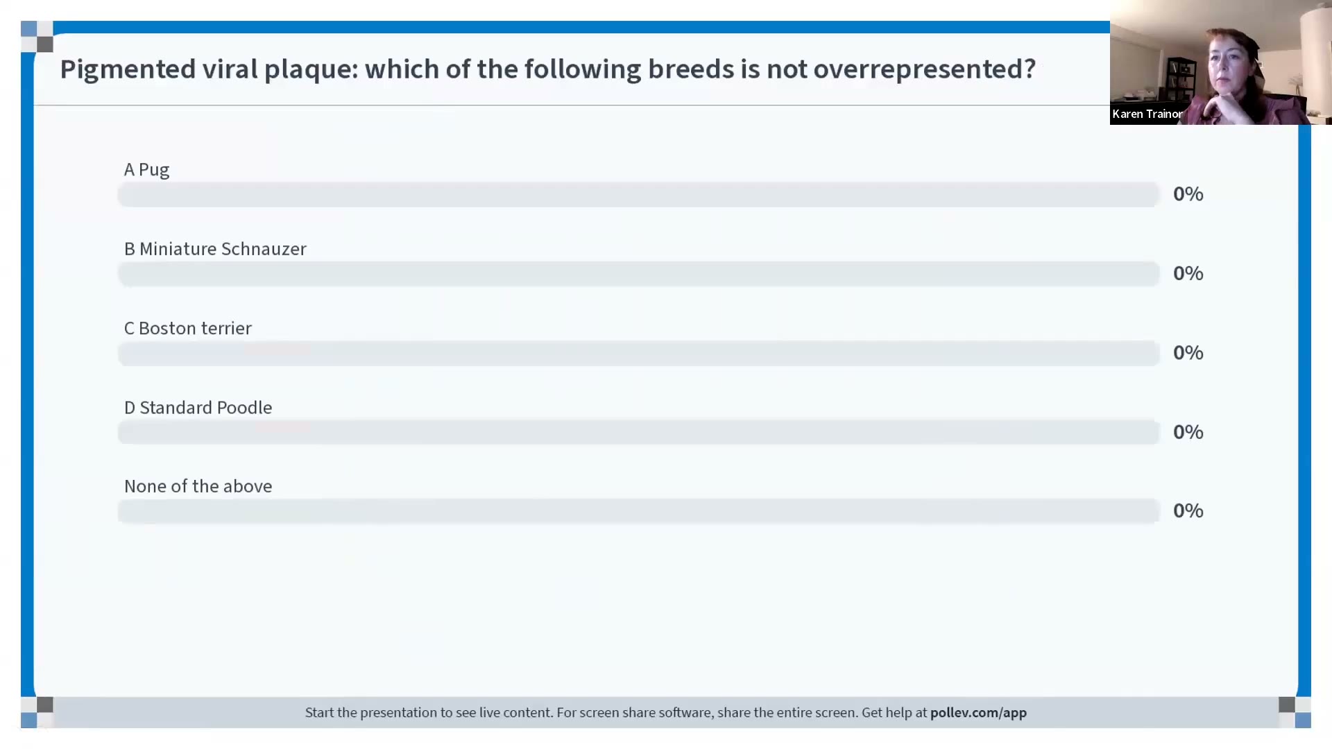
- 🧪 本場小考:「下列哪個犬種沒有好發?」→ standard poodle(標準貴賓)。
2. Squamous cell carcinoma(SCC,鱗狀上皮細胞癌)⏱ 07:45–16:36
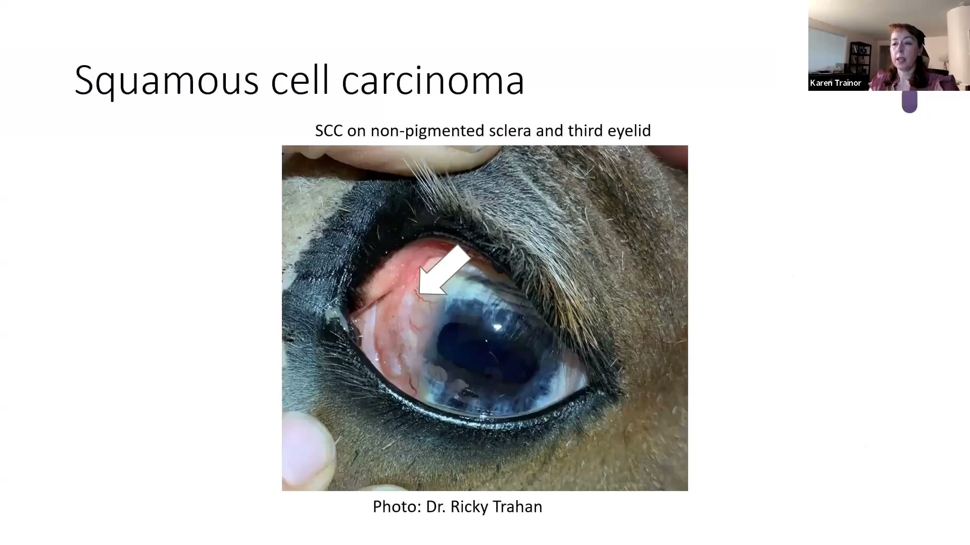
- Clinical:外觀非常多變(pleomorphic) —— 斑塊、火山口狀潰瘍、乳突狀或蕈狀(fungiform)腫塊;有毛皮膚上可脫毛、紅斑、潰瘍或結痂。
- 馬:未著色的鞏膜與第三眼瞼(範例是 paint horse 花馬)
- 牛:「cancer eye(癌眼)」—— Hereford 牛(白臉、眼周白色),尤其日照強的地區(德州)、放牧而非圈養
- 貓:白貓的耳翼、鼻鏡、眼瞼;可先有 **actinic keratosis(日光性角化症)**再進展成 SCC
- 狗:白色短毛犬的背部(或腹側),常曬太陽或住高海拔;別忘了 subungual SCC(甲床下 SCC)
- Cytology:非典型上皮細胞,可見分裂相或怪異細胞核。
- Histopathology:
- 腫瘤細胞穿過基底膜往下浸潤,小葉排列紊亂(disorganized lobules)
- 經典特徵 keratin pearl(角化珠) —— 她推測考試一定會給有 keratin pearl 的圖
- Solar elastosis(日光性彈性組織變性):真皮裡藍灰色的物質=紫外線(UV)傷害 → 「這是原因,這是結果」
- 核仁非常明顯(她形容「從玻片上瞪著你看」)、分裂相多
- 常伴 neutrophilic inflammation,可見 emperipolesis(細胞穿入另一個細胞)
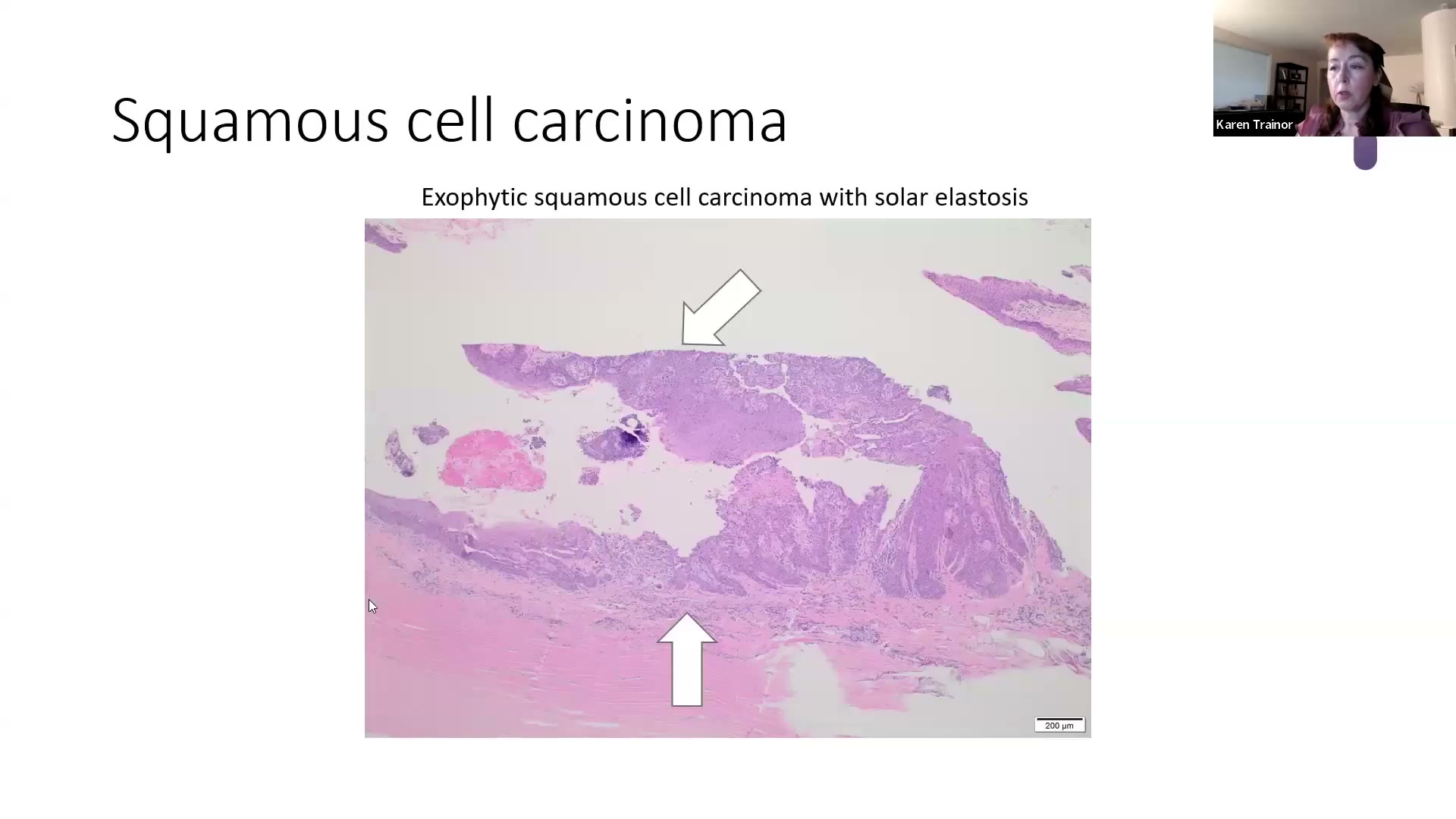
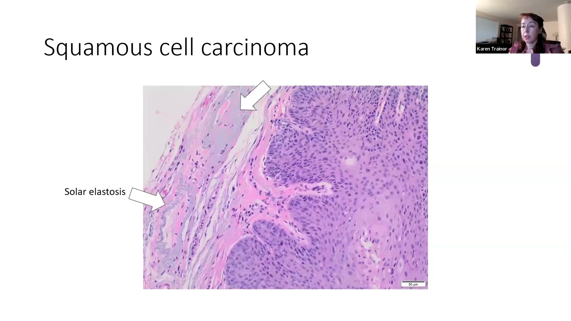
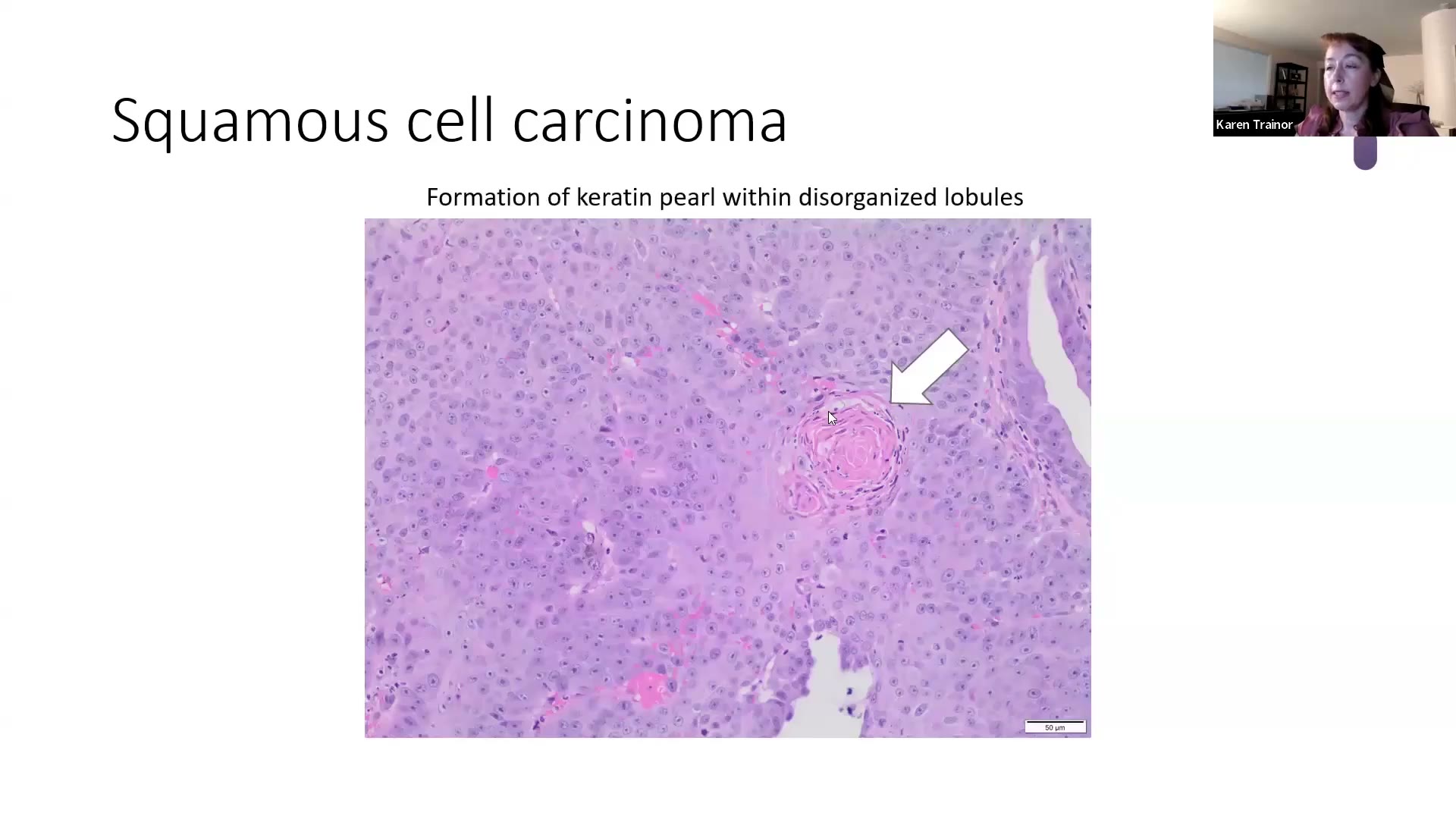
💡 Trainor 的考點
- 🎯 命題老招:把常見病放在「罕見物種」(她舉 milk snake 奶蛇、山羊)—— 「有皮膚的幾乎都會長 SCC」。
- SCC vs IKA(infundibular keratinizing acanthoma,漏斗部角化棘皮瘤):兩者都可以有角化珠/子囊腫
IKA(良性、狗很常見) SCC 小葉 周邊排列整齊 排列紊亂 核仁 不明顯 大而明顯 分裂相 很少 容易找到
- Emperipolesis:neutrophils 穿過腫瘤細胞胞漿(在空泡裡)再從另一邊出來。她看到上皮性腫瘤有這個現象,就「先當 SCC,除非證明不是」,會非常仔細找惡性證據。細胞學或組織切片都看得到。
- UV 相關的腫瘤不只 SCC:淺色短毛犬的腹側、鼠蹊、大腿內側也會出現 solar-induced hemangioma/hemangiosarcoma(日光誘發血管瘤/血管肉瘤),但她說 solar elastosis 比較常跟 SCC 一起看到。
- 大動物重點一句話:Hereford 牛的 cancer eye、paint horse(德州多)。
3. Bowenoid in situ carcinoma(BISC,鮑恩樣原位癌)⏱ 16:36–22:50
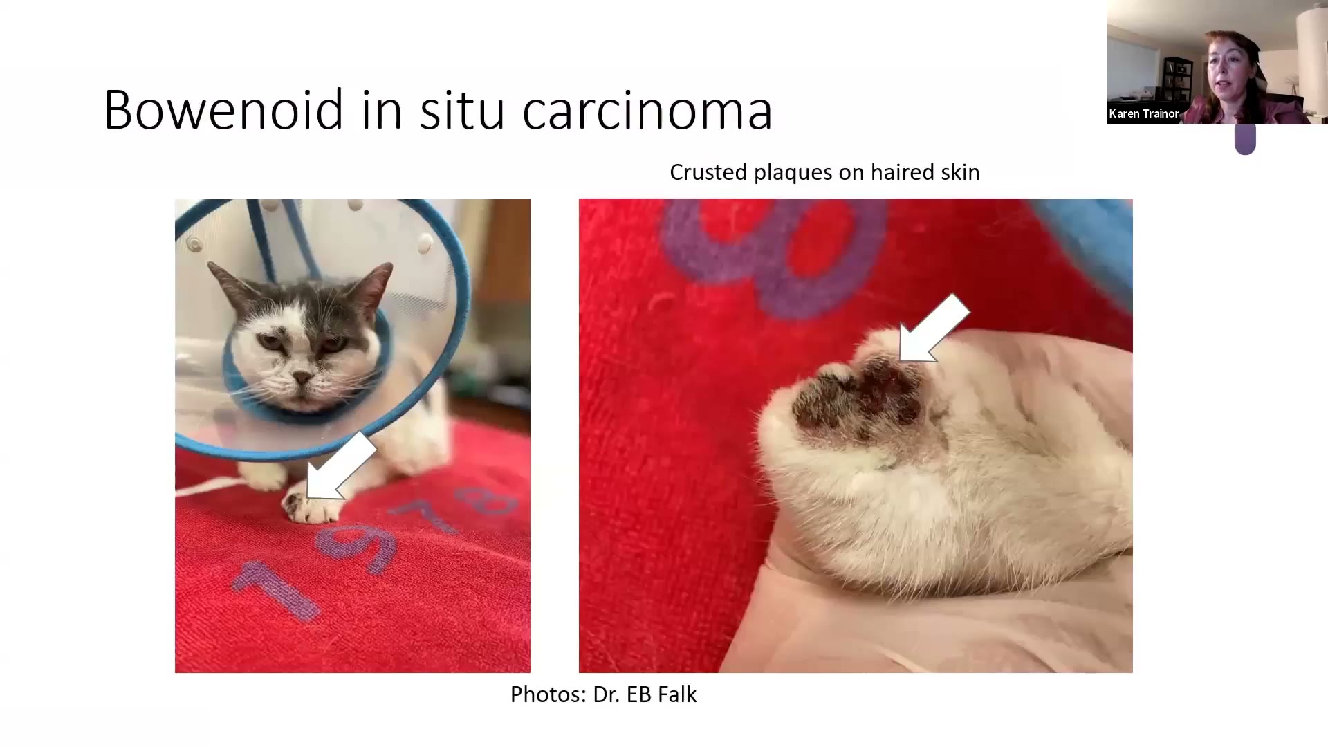
- Clinical:幾乎都是貓;至少 10 歲以上(她看到的多半接近 14 歲)。身上任何部位的多發性、色素沉著、結痂斑塊;著色與未著色皮膚都會長,即使長在白色皮膚上,外觀也可能是黑色斑塊。
- Cytology:可見非典型上皮細胞,但無法評估是否穿過基底膜 → 分不出 in situ 或 invasive,所以要靠組織切片。
- Histopathology:
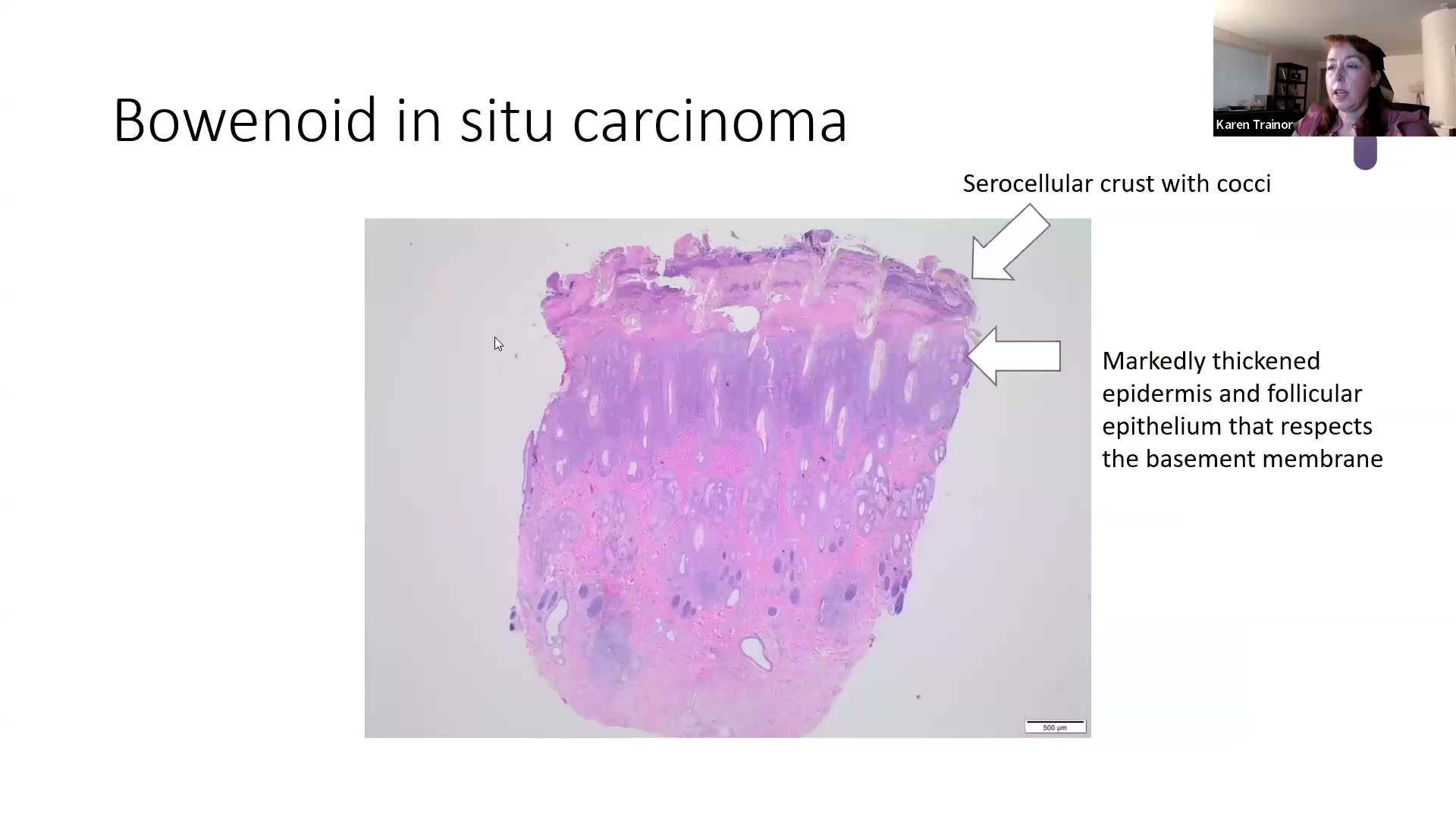
- Serocellular crust,表面常有 cocci 菌落(續發性細菌性膿皮症很常見)
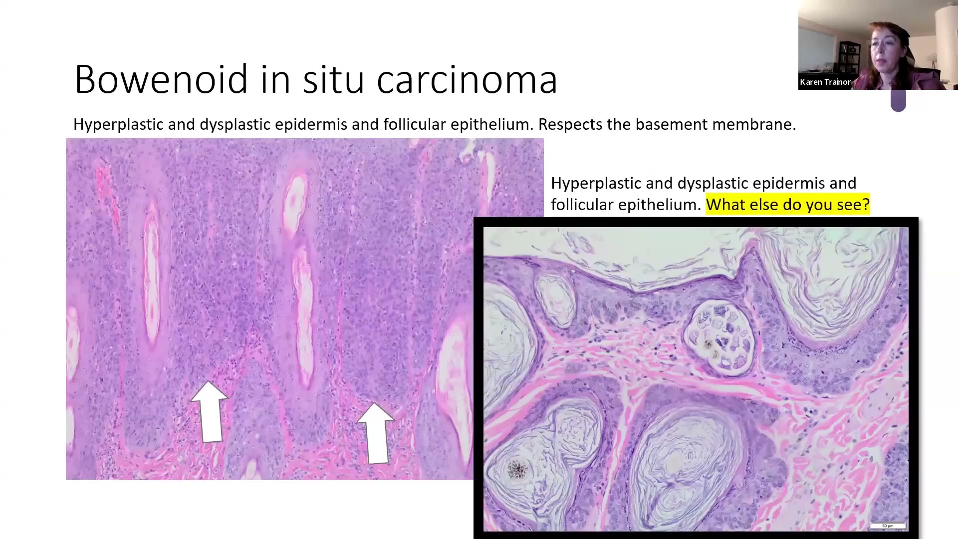
- 表皮與毛囊上皮明顯增厚、增生且異生(dysplastic)
- 🎯 尊重基底膜(respects the basement membrane) —— 會把基底膜往下推、鼓起來,但不突破 → 這就是「in situ」
- DEJ 扇貝狀、表面 hyperkeratosis、granular cell layer 可明顯;表皮與毛囊上皮內常有黑色素
- 看不到病毒包涵體;偶爾胞漿有藍灰色調(viral cytopathic change)
- Additional:與 Felis catus papillomavirus 2(FcaPV-2)最相關。部分貓有在用免疫調節藥物。
💡 Trainor 的考點與臨床提醒
- 惡性轉化比例:投影片寫約 17% 會發展成 invasive SCC 或 basal cell carcinoma(基底細胞癌);她口頭說文獻約 14–15%、另一處說 17–20%。她自己看到的接近 30–40% —— 推測是因為她的病例多來自皮膚科轉診醫院,病灶拖得比較久。→ 考試記 17%。
- 切片要切多處(多發病灶、有的已經侵入)。
- 常合併其他東西:課堂上請學員找圖中毛囊裡的東西 → Demodex;也可合併 Malassezia 過度增生,她甚至看過合併 **pemphigus foliaceus(落葉型天疱瘡)**的病例。
- 發音小插曲:她念「in situ」而不念「in sit-you」,因為後者聽起來像「inside you」。
4. Trichoblastoma(毛母細胞瘤)⏱ 22:50–29:46
- Clinical:非常常見的良性腫瘤;多為單發、脫毛的結節,直徑 1–2 cm。狗多在 6–9 歲。
- 🎯 狗耳根(耳道基部)的下垂、晃來晃去的腫塊 → 幾乎都是 trichoblastoma(她說沒看過那個位置、那種描述是別的東西)
- 貓的常常有色素 → 送檢單上常寫「擔心 melanoma」
- Cytology:「a bag of nuclei(一袋細胞核)」—— 緻密的基底細胞團塊;細胞學上歸在 basal cell tumor 一類(少數 apocrine gland 腫瘤也長這樣)。可以有囊腫性退化:抽到上皮細胞加一堆液體時要想到 cystic trichoblastoma。
- Histopathology:邊界清楚、細胞密集
- **狗=ribbon type(緞帶型)**最常見,也可形成 medusoid(梅杜莎頭狀)漩渦
- 貓=trabecular type(小樑型) —— 細胞沿小葉周邊整齊列隊,很像口腔的 ameloblastoma(造釉細胞瘤)
- Additional:還有其他亞型(如 granular cell type),考皮膚科專科不需要管。
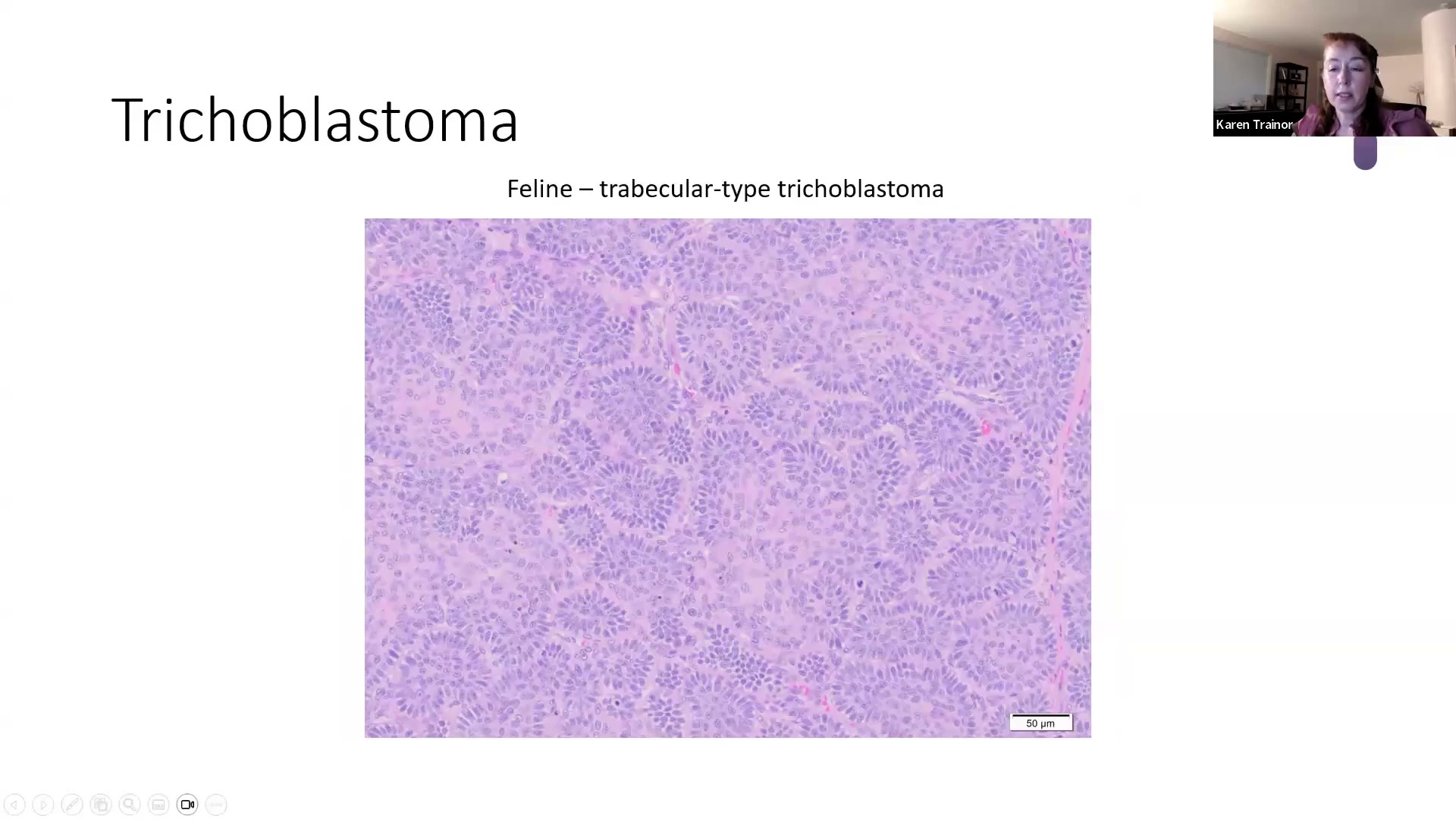
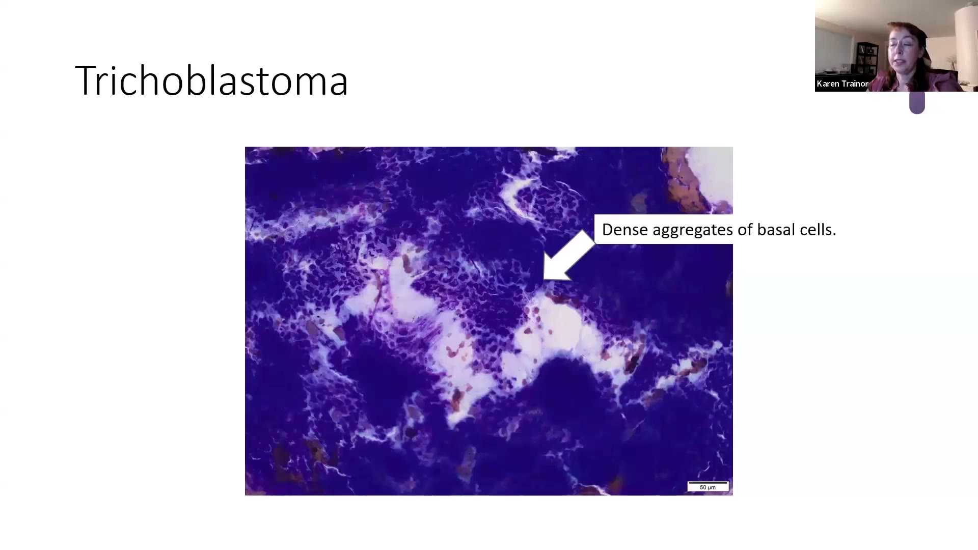
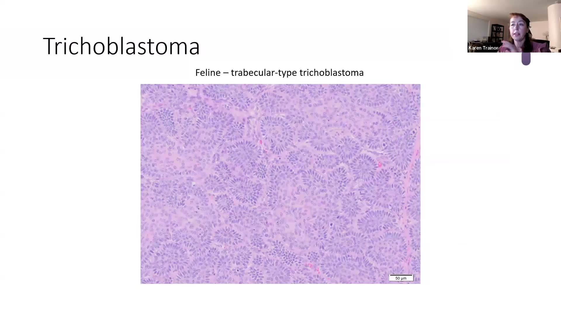
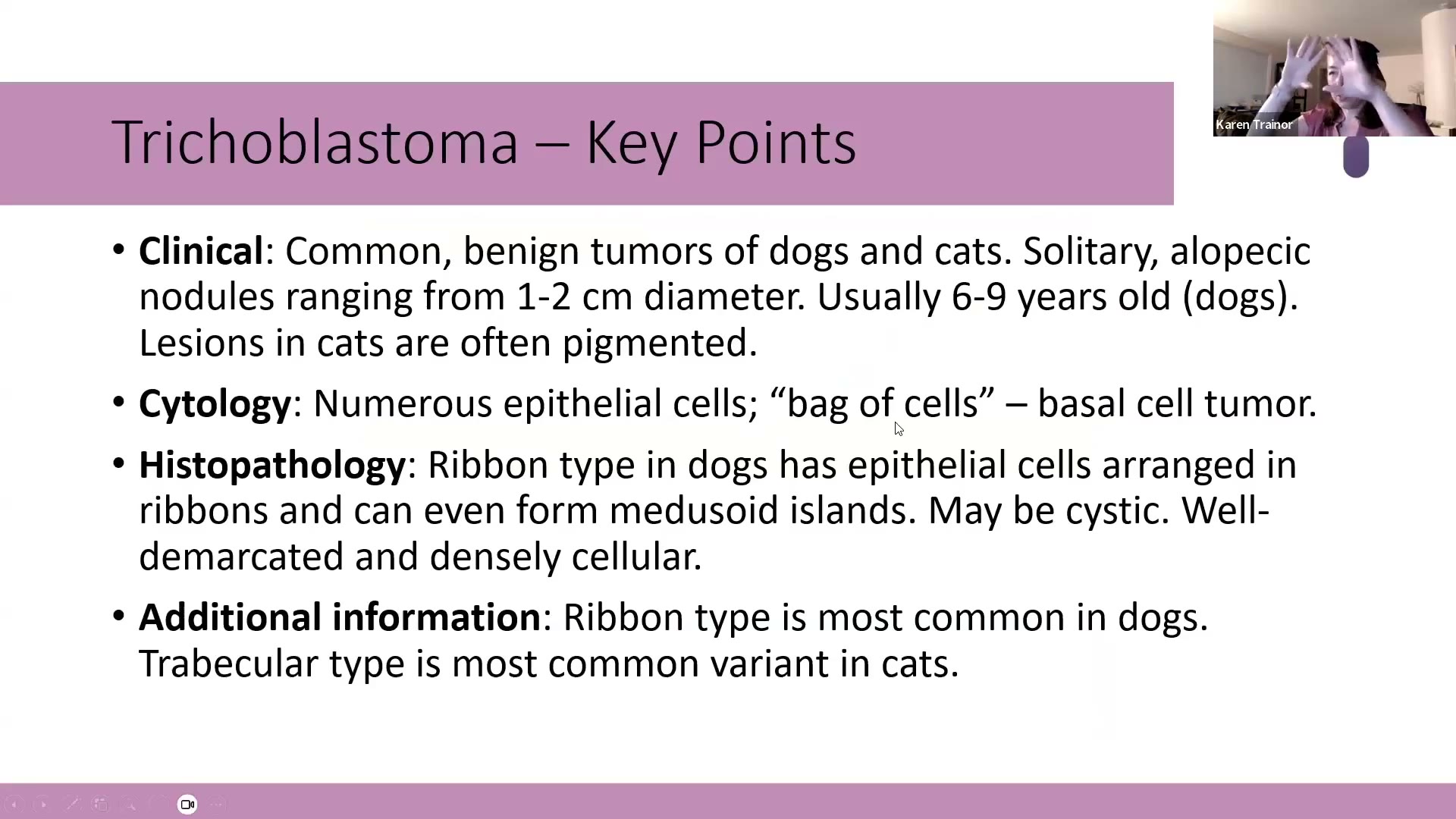
💡 Trainor 的考點
- 如果只能背一個毛囊腫瘤,就背 canine ribbon-type trichoblastoma:常見、細胞學漂亮、組織病理經典、幾乎沒有別的東西長這樣、沒有爭議。
- 她自己看了幾十到上百例才掌握各種變化 → 建議多看圖。
- 🎯 反向出題的可能:給你 trabecular trichoblastoma 的圖,問物種 → 答「貓」。她說以前病理專科考試只寫「tissue from a ___」,要你自己填完整描述與診斷。她不認為皮膚科專科會這麼刁鑽,但要熟到不管題目給哪幾塊拼圖都答得出來。
- 臨床:耳根下垂腫塊可以細胞學確認後,有造成困擾再切除。
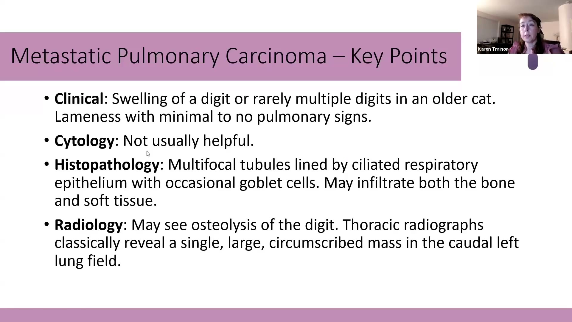
5. Metastatic pulmonary carcinoma(肺癌轉移至趾,lung-digit syndrome)⏱ 29:46–34:50
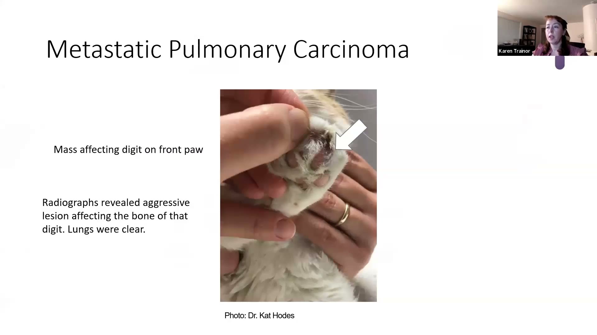
- 名稱:metastatic pulmonary carcinoma 或 metastatic pulmonary adenocarcinoma(她說看你要多精確)。
- Clinical:老貓(約 12 歲以上)前腳單一趾頭腫脹,罕見多趾;可伴跛行;奇怪的是呼吸道症狀很少或沒有。
- 範例貓:前腳一趾腫塊,X 光顯示該趾骨頭侵襲性病灶,肺部卻是乾淨的(她沒拿到放射科報告,不確定有沒有給放射科醫師看)
- Cytology:以前認為不太會脫落;她當場更正:有同事剛分享過細胞學成功的病例 → 可以試,但直接截趾送組織病理可能是最好的診斷方式。
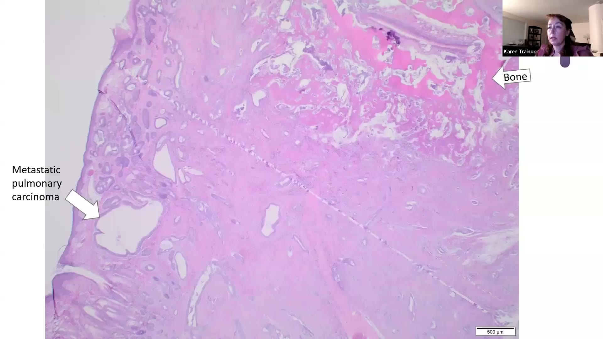
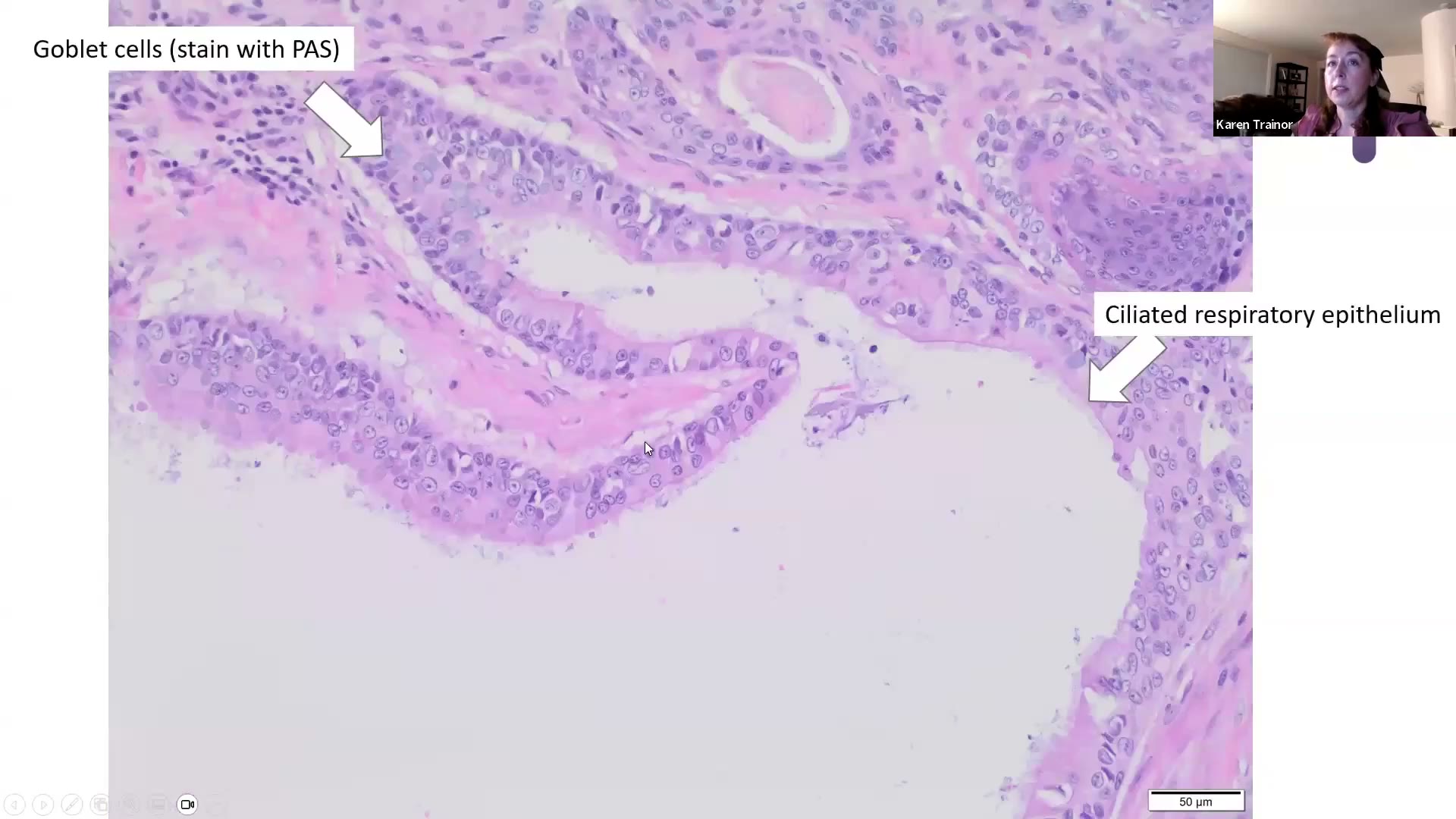
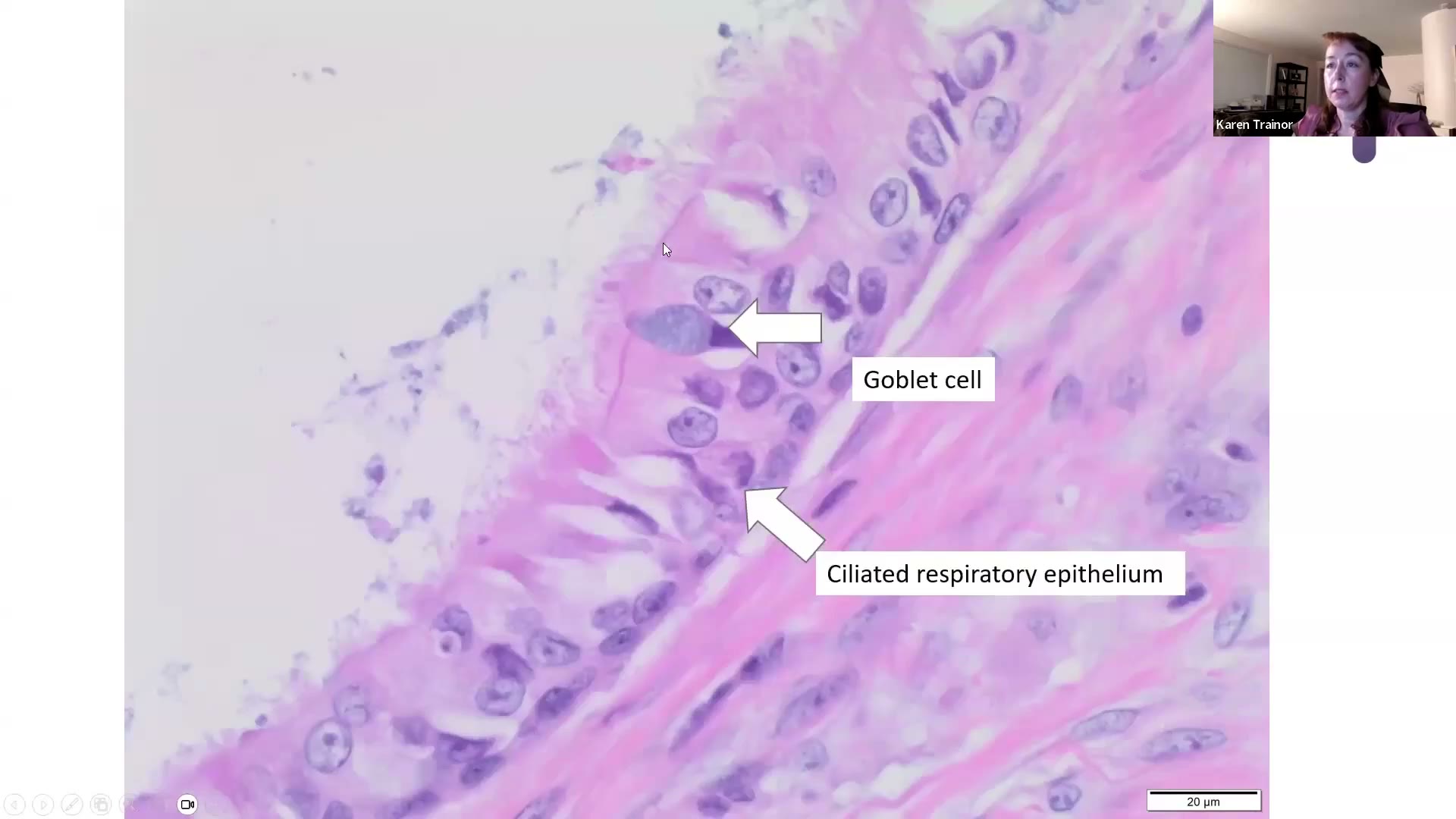
- Histopathology:
- 多發的小管或囊腔,內襯 ciliated respiratory epithelium(纖毛呼吸道上皮)+ goblet cells(杯狀細胞)
- 🎯 找亮粉紅色的線=纖毛附著處(mucociliary apparatus)→ 這就是下診斷的依據
- 杯狀細胞胞漿內藍灰色的是 mucin → 可用 PAS(periodic acid-Schiff)或 Alcian blue 染
- 腫瘤同時侵犯骨頭與軟組織,伴骨膜新生骨;細胞核開始**堆疊(pile up)**是惡性表現
- Additional:
- 趾頭 X 光:趾骨溶骨(osteolysis)
- 胸腔 X 光:經典教科書描述是單一、界線清楚的大腫塊,位在「左後肺野(caudal left lung field)」(她說自己的病例不常這樣,但考試應該會照書出)
💡 Trainor 的臨床提醒
- 老貓只有一趾腫 → 趾頭與胸腔都要照 X 光。
- 範例切片有刀痕(chatter)假影,因為帶骨頭的切片很難切。
6. Equine sarcoid(馬類肉瘤)⏱ 34:50–41:36
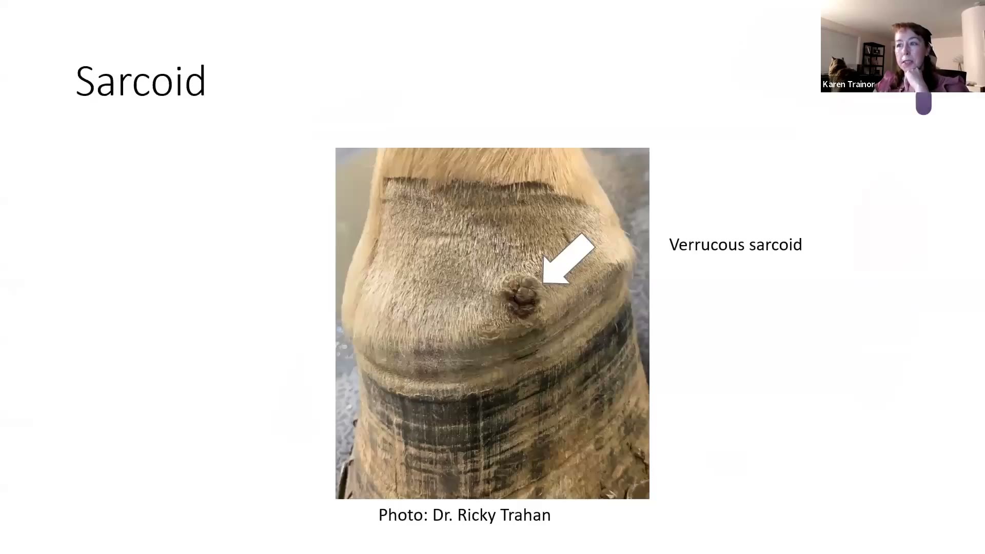
- Clinical:馬、驢、騾、斑馬最常見的皮膚腫瘤 —— 高達 90% 的馬科皮膚腫瘤是 sarcoid。好發頭、四肢、腹側軀幹。
- 六種亞型:verrucous(疣狀)、flat/occult(扁平/隱匿)、nodular(結節)、fibroblastic(纖維母細胞型,像 proud flesh 肉芽組織過度增生)、mixed(混合)、malignant/malevolent(惡性)。她最常看到 verrucous、nodular、fibroblastic、mixed;flat 與 malignant 很少見。
- Cytology:沒有幫助 —— 因為是 biphasic,診斷全靠組織架構。
- Histopathology:
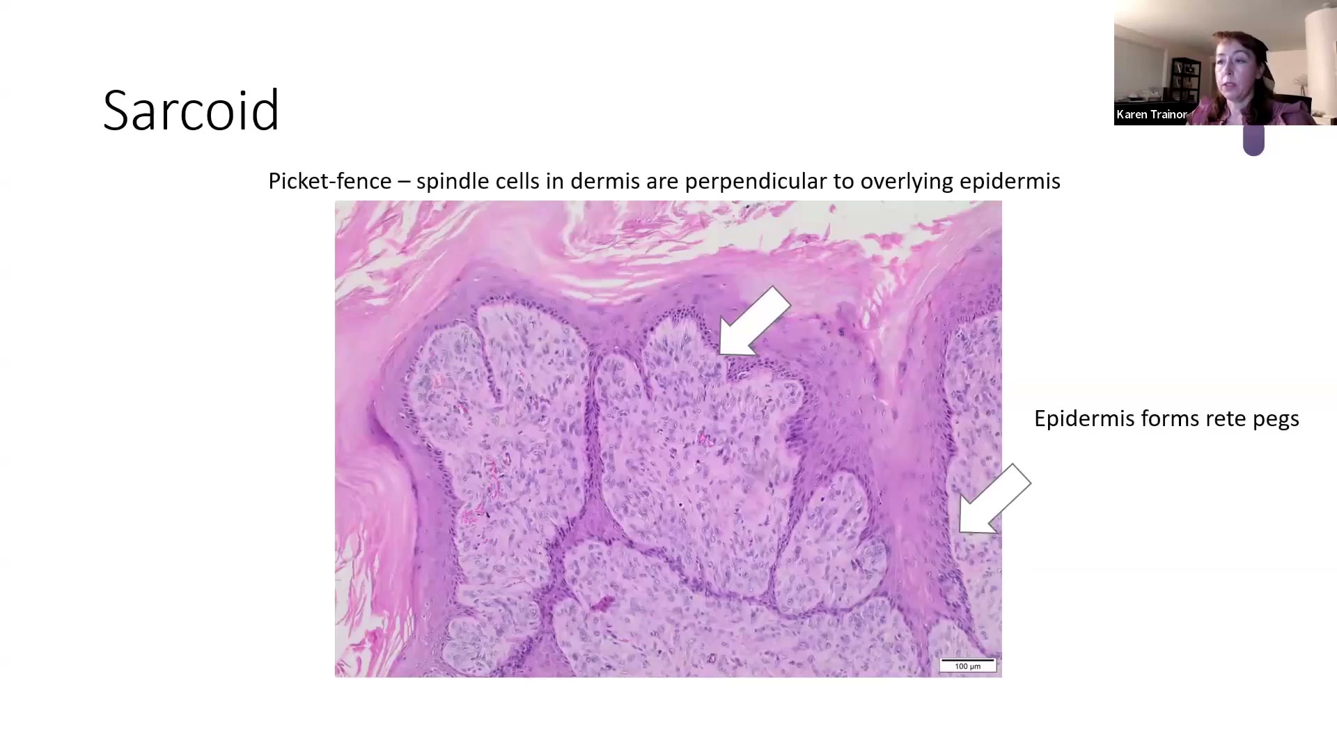
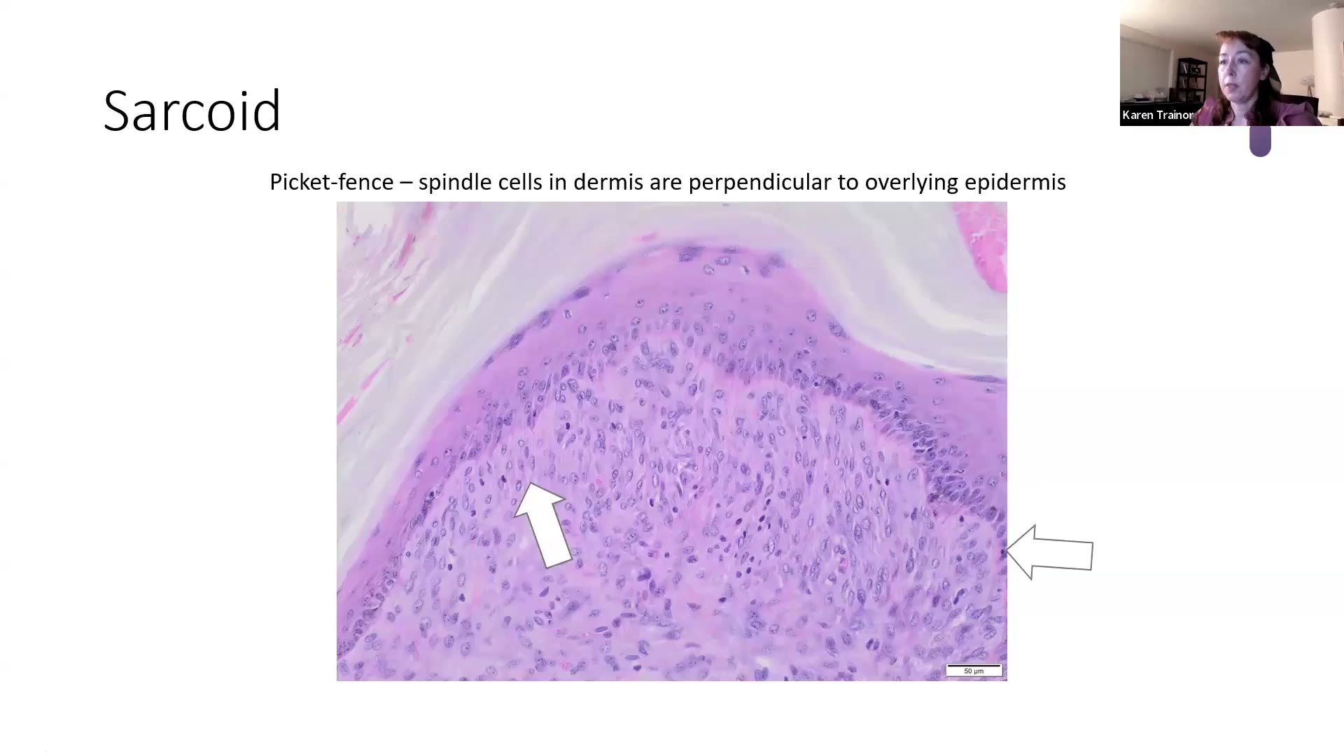
- 🎯 Biphasic(雙相性)腫瘤:表皮成分(pseudoepitheliomatous hyperplasia)+ 真皮的 spindle cell 成分,要看到兩者的交界互動才能確診
- 🎯 Picket fence(尖樁籬笆)pattern:真皮的纖維性 spindle cells 一根根排列,垂直於(perpendicular)表皮基底膜與 rete pegs
- Additional:Bovine papillomavirus(BPV,牛乳突病毒)1、2、13(還有其他幾型),多數 sarcoid 表現 BPV-1 與 BPV-2;主要轉化病毒蛋白是 E5 → 下調 MHC I 表現,讓腫瘤躲過 cytotoxic T cells 的免疫清除。
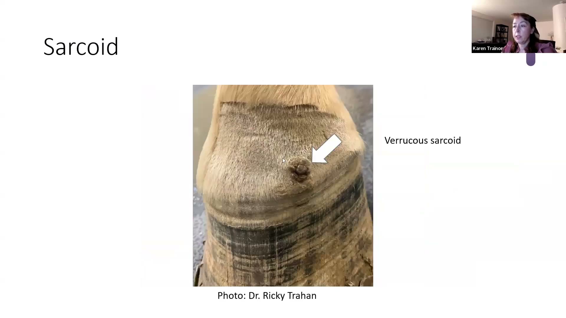
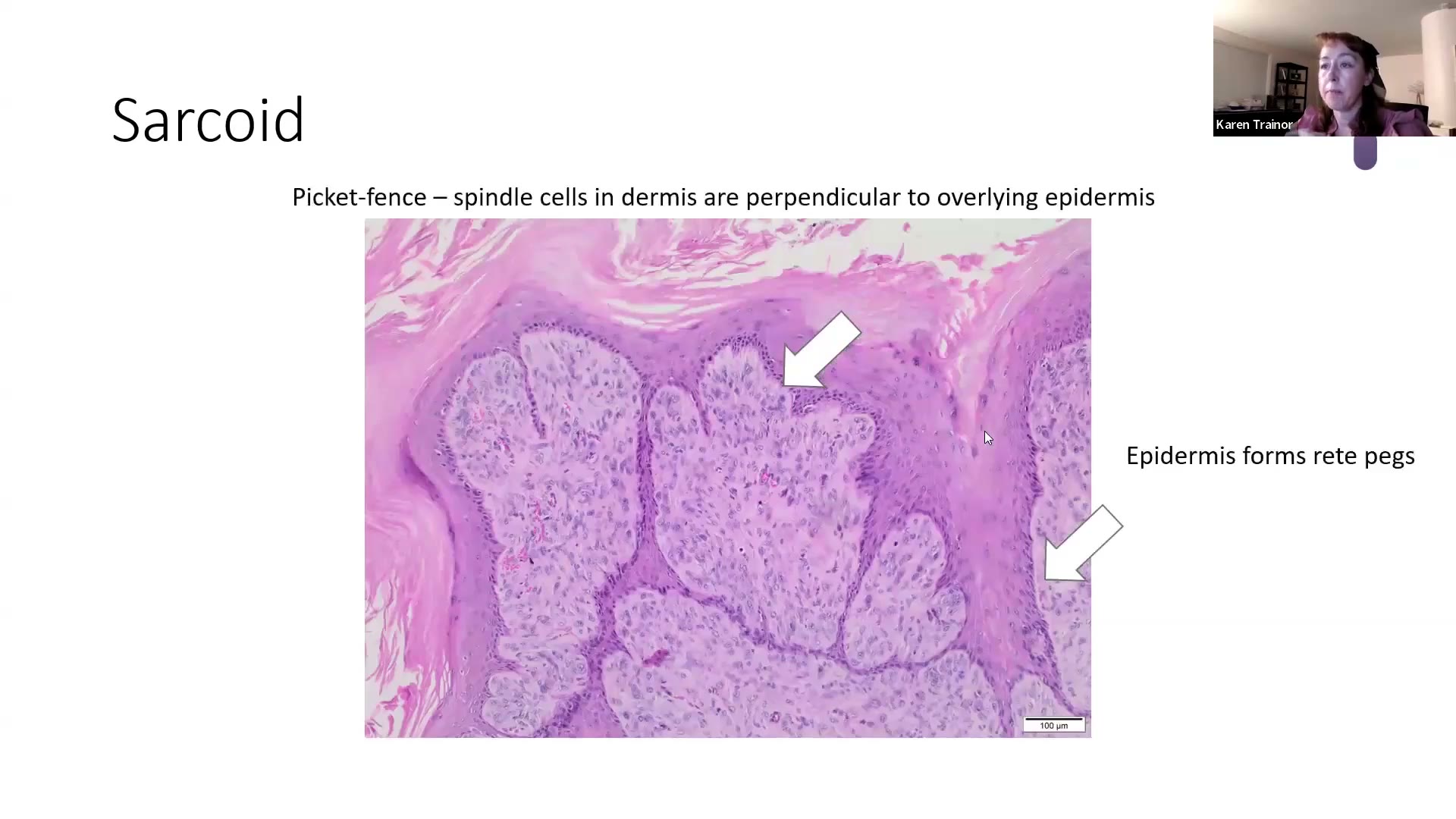
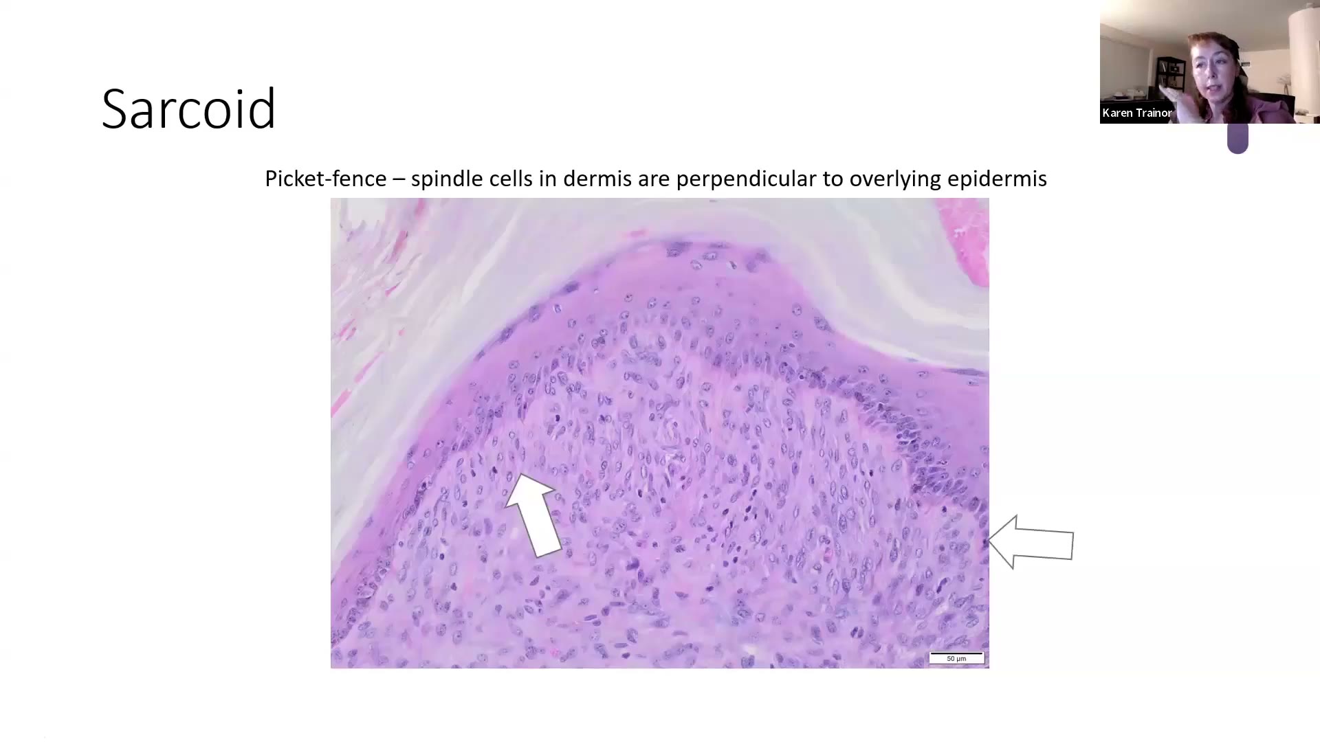
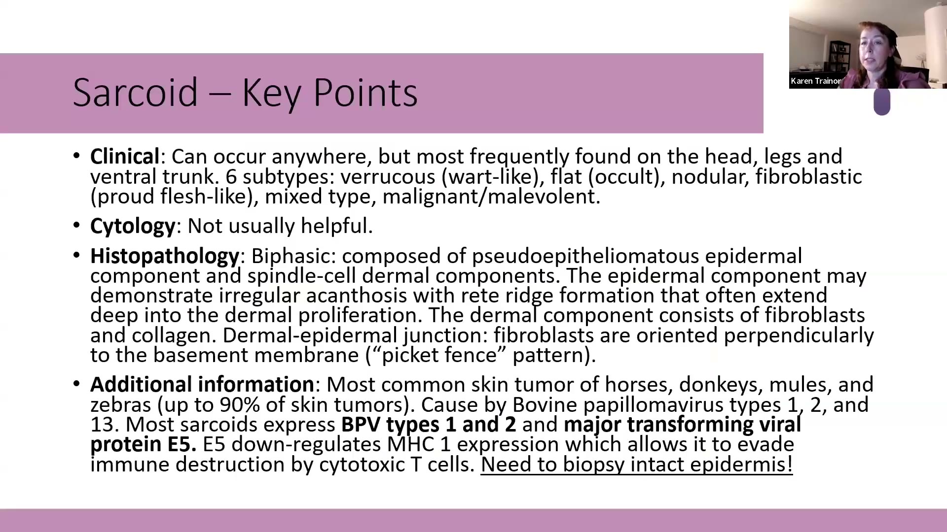
💡 Trainor 的「肥皂箱」(她自己說的,最想讓大家記住的實務重點)
採檢一定要有完整、沒有潰瘍的表皮覆蓋在真皮上。
- 因為是 biphasic,沒有表皮就無法確診 → 病理只能寫「suspect sarcoid」,大家都很不開心。
- 腫塊有潰瘍時,盡一切努力從沒潰瘍的地方取樣。
- 這不只是考點,是實務上要跟所有一般科醫師宣導的事。
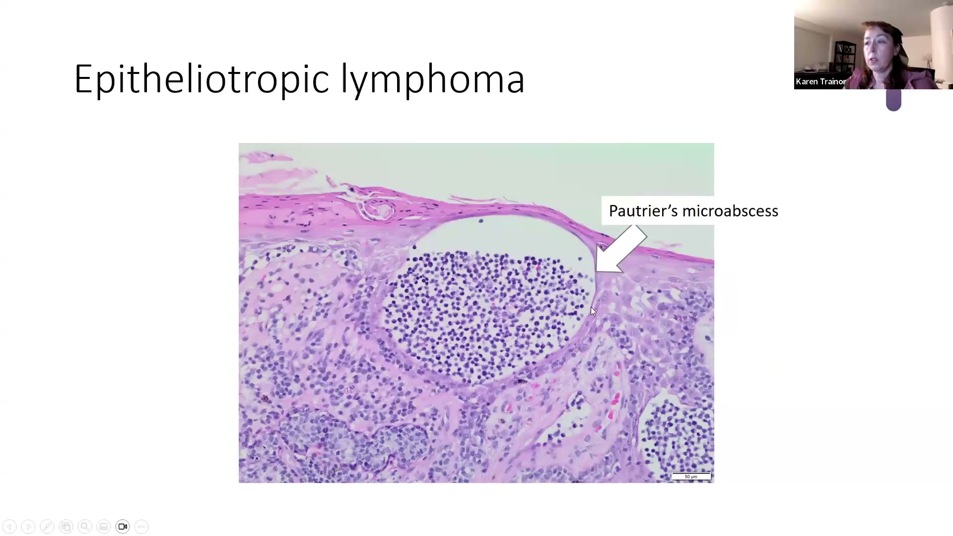
7. Epitheliotropic lymphoma(親上皮性淋巴瘤)⏱ 41:36–46:00
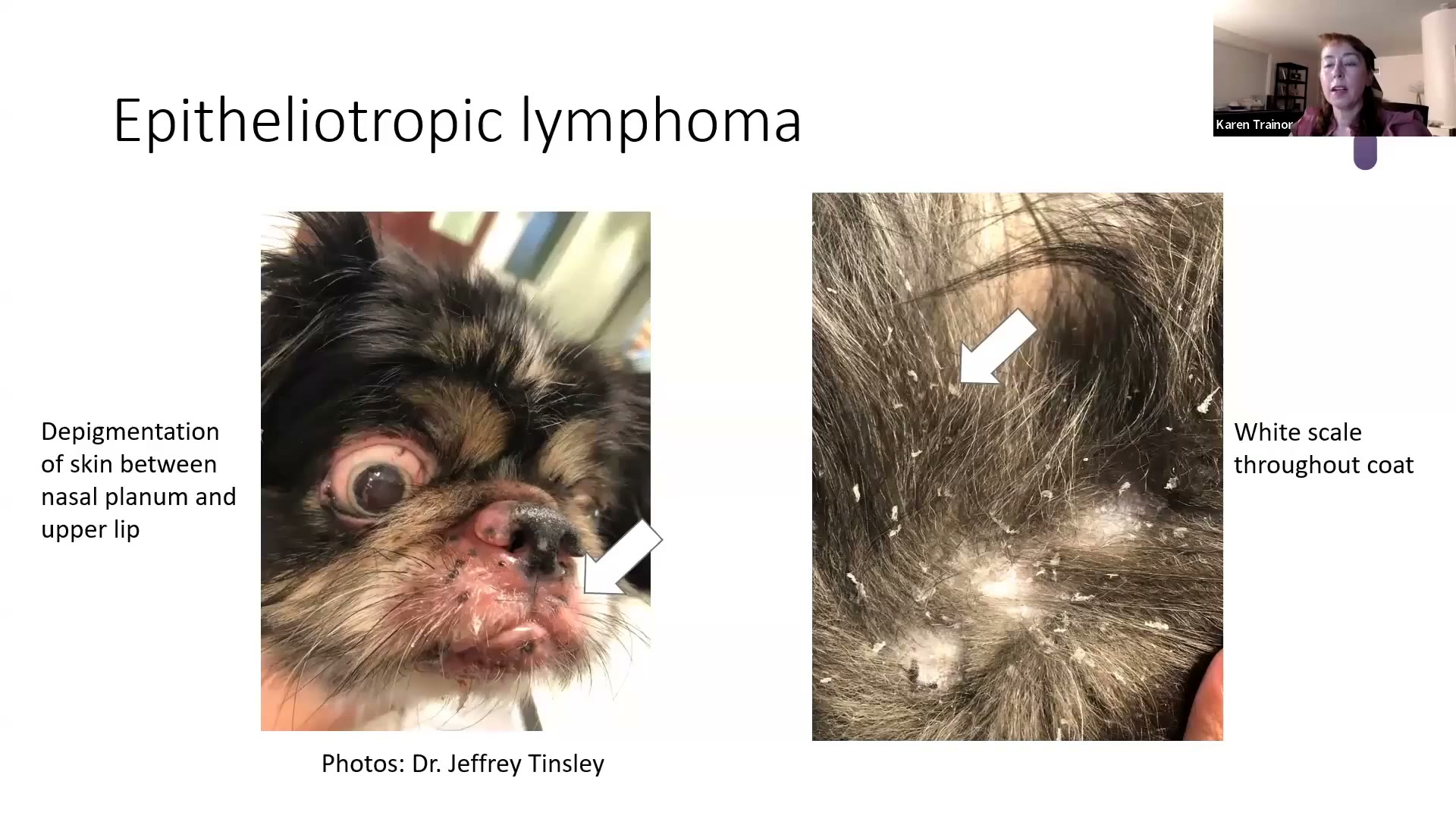
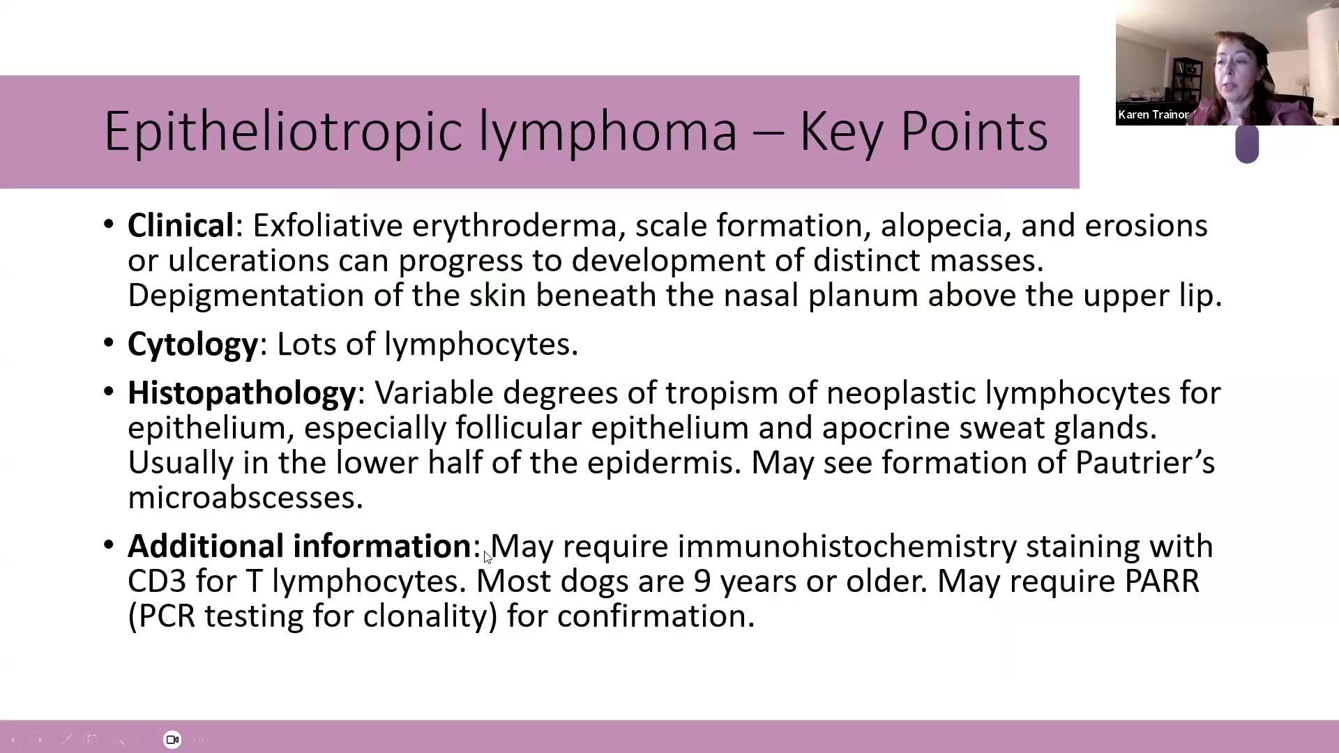
- Clinical:
- 🎯 鼻鏡與上唇之間去色素,鼻鏡、上唇本身也可受影響 → 「八字鬍(mustachioed)外觀」→ 先當淋巴瘤,除非證明不是
- 全身厚的白色、帶銀光的大片皮屑
- 剝落性紅皮症(exfoliative erythroderma)、脫毛、糜爛、潰瘍,之後可能長出明顯腫塊
- 多數 ≥9 歲;範例狗只有 4 歲
- Cytology:大量淋巴球 → 「讓人緊張的時刻」→ 一定要切片確認(要看組織架構)。
- Histopathology:
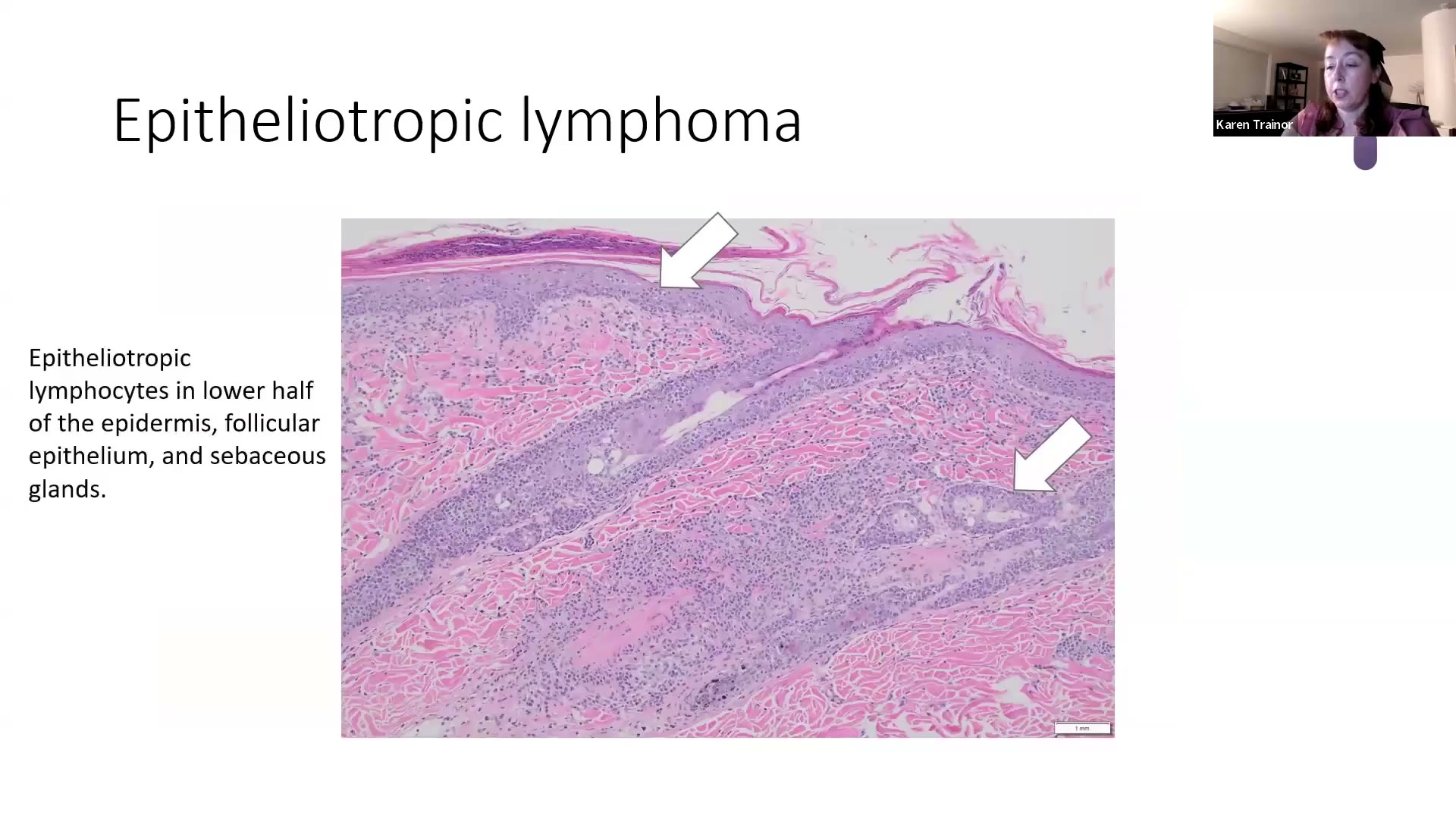
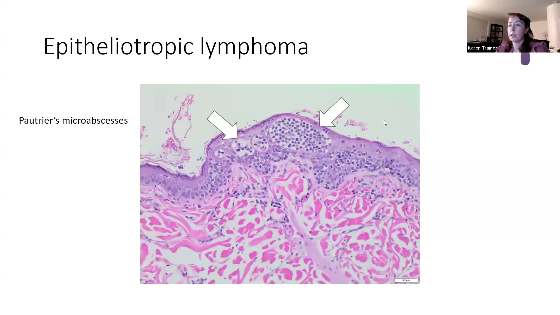
- Epitheliotropism:腫瘤淋巴球浸潤表皮、毛囊上皮、皮脂腺,也可到 apocrine 汗腺
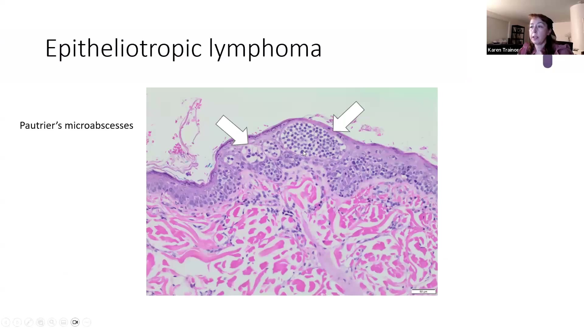
- 🎯 Pautrier microabscesses(Pautrier 微膿瘍) —— 有些病理醫師改稱 Pautrier aggregates(聚集)(因為既不「微」也不是「膿瘍」),但教科書與考題年年用 microabscesses,兩個名詞都要認得
- 腫瘤細胞偏向表皮或毛囊上皮的下半部
- Pagetoid reticulosis:淋巴球只集中在表皮內的型態
- Additional:IHC(immunohistochemistry,免疫組織化學)染 CD3 確認 T 淋巴球;困難病例需做 PARR(PCR for antigen receptor rearrangement,淋巴球抗原受體重組 PCR 檢測) 驗 clonality。
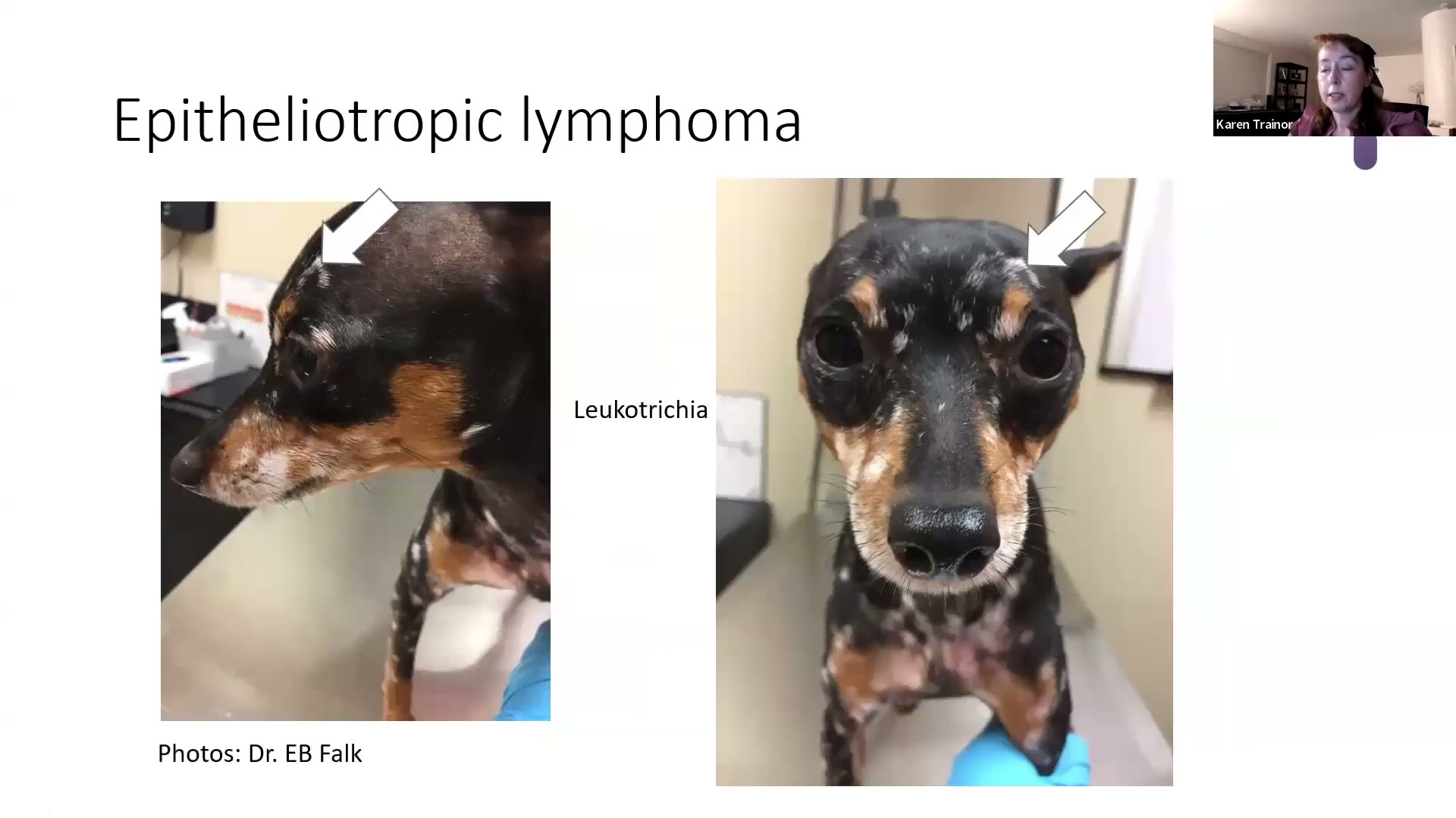
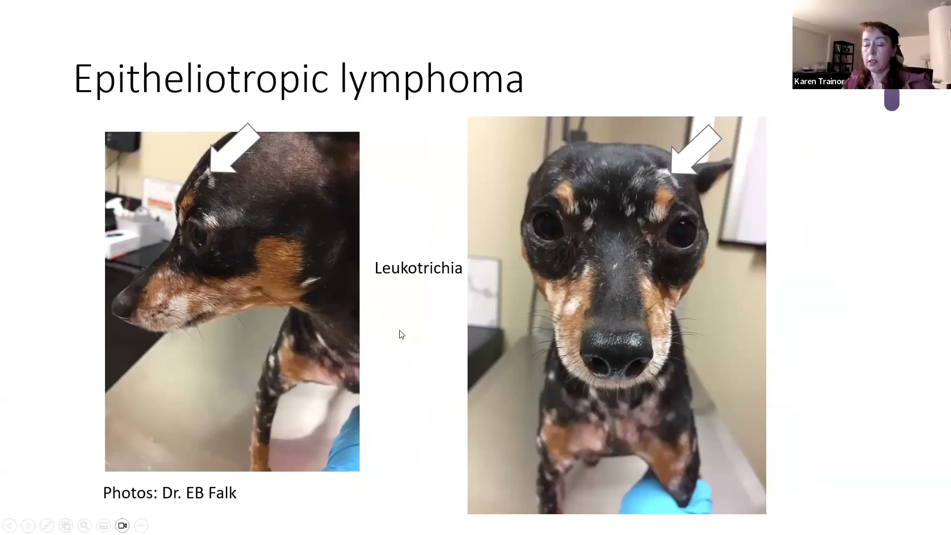
💡 Trainor 的考點與臨床提醒
- 🎯 「我敢打賭考試出這題一定會有 microabscesses」;唯一例外是給你 pagetoid reticulosis 的圖。
- 年輕狗出現 leukotrichia(毛髮變白)也要切,而且多切幾處(leukoderma + leukotrichia 的地方),送皮膚病理醫師 —— 因為會造成 leukotrichia 的病很少,要排除淋巴瘤;也可能只是 vitiligo(白斑症)。
- 她說被淋巴瘤「教訓過很多次」:淋巴瘤什麼樣子都可能出現,她不喜歡太快下淋巴瘤診斷,但一定要檢查。
- 🧪 本場小考:「哪個診斷經典地與 Pautrier microabscesses 相關?」→ epitheliotropic lymphoma。
貳、Tumors 小考(5 題)⏱ 46:00–49:16
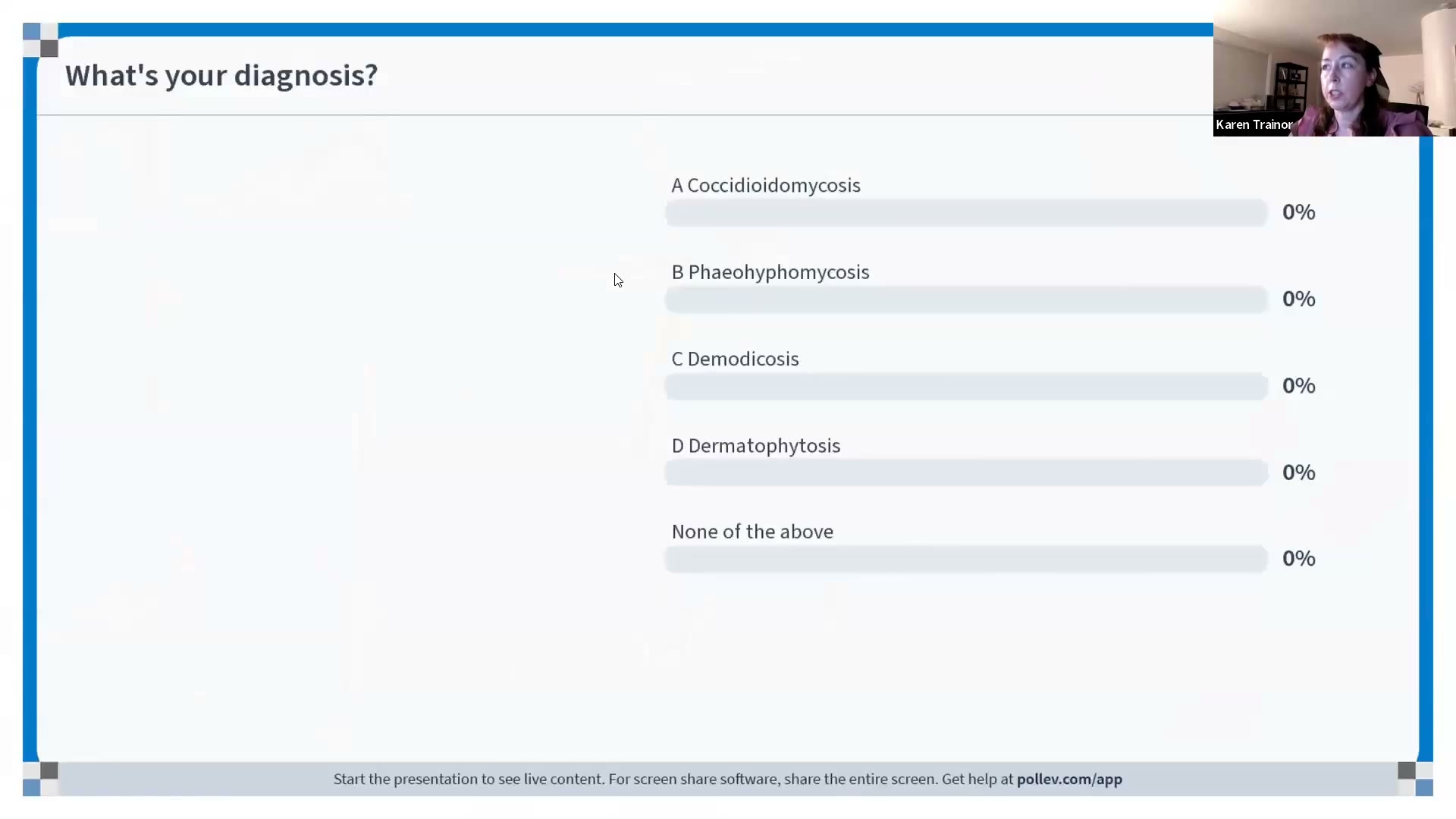
原本用 Pollev QR code 投票,學員看不到圖(技術問題),她改成直接念題目、學員打在 chat。
| # | 題目 | 正解 | 解析/連結 |
|---|---|---|---|
| T-Q1 | What’s your diagnosis?(選項:Coccidioidomycosis/Phaeohyphomycosis/Demodicosis/Dermatophytosis) | Dermatophytosis | 染色淡的菌絲+arthrospores 圍在毛幹周圍(chat 有人答 Demodex,因為看不到圖)→ 2026.08.28 Board Review Part 2 > 13. Dermatophytosis(皮癬菌症)⏱ 80:52–86:50 |
| T-Q2 | Alopecia areata 的經典組織學發現? | Bulbitis —「swarm of bees」 | → 2026.08.28 Board Review Part 2 > 16. Alopecia areata(圓禿)⏱ 97:57–102:53 |
| T-Q3 | 犬 Cushing’s(hyperglucocorticism)的特徵是 thin-walled comedones、表皮萎縮、皮脂腺萎縮? | True | → 2026.08.28 Board Review Part 2 > 17. Canine Cushing’s disease(犬庫欣氏症)⏱ 103:07–107:03 |
| T-Q4 | Pigmented viral plaque:哪個犬種沒有好發?(pug/miniature schnauzer/Boston terrier/standard poodle/none) | Standard poodle | 見本筆記腫瘤 1 |
| T-Q5 | 哪個診斷經典地與 Pautrier’s microabscesses 相關?(melanocytoma/metastatic pulmonary adenocarcinoma/sarcoid/epitheliotropic lymphoma) | Epitheliotropic lymphoma | 見本筆記腫瘤 7 |
小考投影片


參、Q&A ⏱ 49:16–52:12、62:16–64:32
現場 沒有人提問
她開放 chat 等了一兩分鐘,沒有問題,就直接進 mock exam;最後又開放一次,還是沒有。 → 您先前列的 5 個問題沒有在這場被回答,要問的話直接寄信:karen@innovativevetpath.com(她說看錄影後才想到的問題、考前任何問題都歡迎寄)。
她在 Q&A 時段交代的事:
- 考試形式:mock exam 刻意只用選擇題、不計時、不用 QR code。
- 課後問卷:她想知道 ① 8–9 月上課時間太早或太晚?② 資訊量太多或不夠?③ 想不想要其他主題的考前複習(生理、藥理、寄生蟲),時間排在 11 月考前(因為 Resident Education Forum 跟考試時間剛好相反)。
- 錄影開放到考試前(課程頁寫 2026/11/15)。
- 10 月開 DermPath 101:拆解基礎、以詞彙為主、更多複習,一樣 Elanco 贊助。
- 記得感謝 Elanco 與業務代表的贊助。
- 她為 Part 2 結尾的技術問題道歉。
- Q&A 開場前放了一張改編〈聖誕夜前一晚〉的考前打油詩(‘Twas the night before boards…),最後一句是住院醫師夢裡都是 Demodex。
肆、Mock Exam(選擇題)⏱ 52:12–62:16
這份 mock exam 全部出自 Part 1(表皮疾病),題目幾乎就是 Part 1 各病的 Key Points 換句話說 → 答錯的題目直接回 Part 1 對應段落重讀。
她在台上的評語:「You guys are on fire」—— 全場幾乎都答對,所以這些是「基本盤」,考試不能錯。
Q1 ⏱ 52:12 — 貓的 pemphigus foliaceus(PF,落葉型天疱瘡)病灶最常出現在哪?
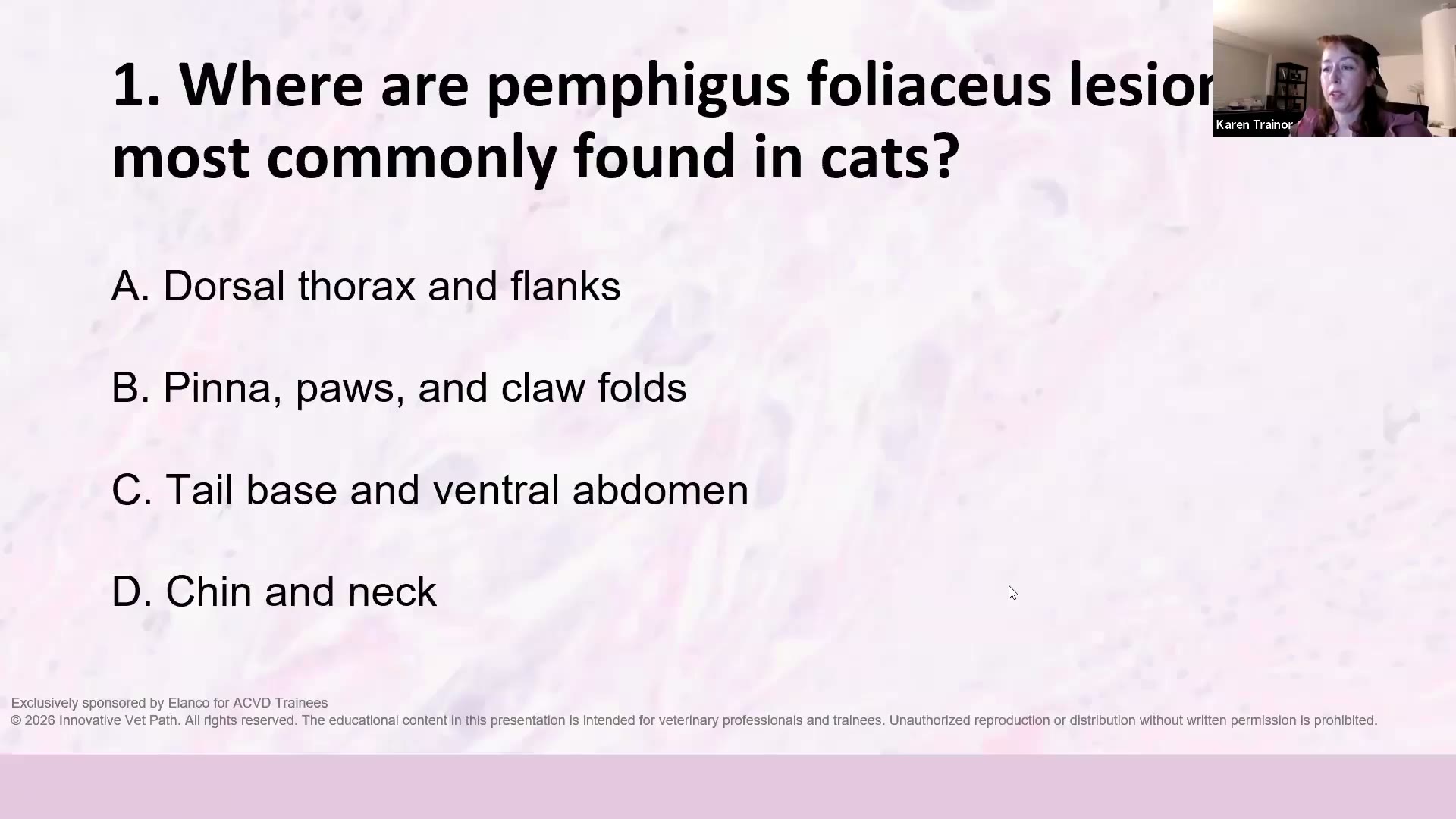 (A) Dorsal thorax and flanks (B) Pinna, paws, and claw folds ✅ (C) Tail base and ventral abdomen (D) Chin and neck
解析:耳翼、腳掌、爪褶。她說:不只自己要知道,也要讓轉診醫院的醫師和新手獸醫都知道。 → Part 1:1. Pemphigus foliaceus
Q2 ⏱ 53:10 — PF 經典的組織學 pattern?
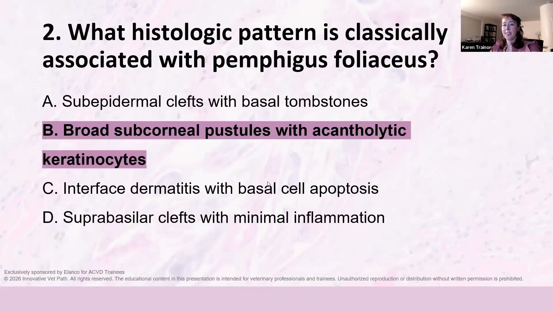 (A) Subepidermal clefts with basal tombstones (B) Broad subcorneal pustules with acantholytic keratinocytes ✅ (C) Interface dermatitis with basal cell apoptosis (D) Suprabasilar clefts with minimal inflammation
解析:寬大的角質層下膿疱+棘層鬆解角質細胞(acantholytic keratinocytes)。其他選項:
- D「suprabasilar clefts」= PV 的裂開層次。Gross 2005 與 Muller & Kirk 7e:PV 在基底上裂開,bulla 頂部脫落後,一列基底細胞仍黏在基底膜上=tombstone(tombstone 是基底上裂開的結果)。基底上裂開的還有 PNP(有凋亡)、Darier’s(全層崩解磚牆、IF 陰性)。D 錯在層次(PF 在表皮上層)和發炎程度(PF 是膿疱,不是發炎很少)。⚠️ vault 裡的課本筆記沒寫 PV 的發炎程度,Trainor 說 PV 裂隙內有出血與 serocellular exudate → 「minimal inflammation」應是誘答的拼湊。
- A「subepidermal clefts with basal tombstones」自相矛盾:表皮下裂開時,基底細胞會跟著頂部被掀走,地板只剩基底膜(JEB 用 PAS 可見 lamina densa),不會有墓碑。
- C 是 interface(細胞毒殺型)pattern,例如 EM、lupus。

課本:尋常性天疱瘡 Pemphigus vulgaris|MK7e 09-06 Pemphigus Vulgaris|犬 Darier 氏病 Canine Darier’s disease
📖 原文:JEB(Hereditary junctional epidermolysis bullosa)
Gross et al. 2005, Skin Diseases of the Dog and Cat 2e, pp. 38–40(Histopathology) “In both junctional and dystrophic EB, the epidermis is cleanly separated from the underlying dermis to form a bulla or vesicle. Inflammation is not present within early bullae, and dermal inflammation is sparse.” “A periodic acid–Schiff (PAS) stain may color the lamina densa along the floor of the bulla in junctional EB and on the roof of the bulla in the dystrophic form.” “Conversely to dystrophic EB, immunomapping showed the separation above the lamina densa in dogs with junctional EB, indicative of cleavage through the lamina lucida (Olivry et al., 1997).” “Inflammation within and beneath the intact cleft of inherited EB is generally sparse in comparison to the other subepidermal bullous dermatoses, except for some cases of linear IgA disease.” 圖說(Fig. 2.19):“Basal cells remain intact in the roof of the bulla.”
Miller, Griffin & Campbell 2013, Muller & Kirk’s Small Animal Dermatology 7e, Ch.12 “In junctional epidermolysis bullosa, blisters form within the lamina lucida of the dermoepidermal basement membrane.” “Histologically, animals with junctional epidermolysis bullosa show a subepidermal separation with little or no subjacent dermal inflammation.”
💡 重點:基底細胞跟著頂部被掀起 → 地板沒有墓碑;「發炎很少」是課本寫給 JEB/遺傳性 EB 的,不是 PV。
📖 原文:PNP(Paraneoplastic pemphigus)
Gross et al. 2005, Skin Diseases of the Dog and Cat 2e, pp. 36–37(Histopathology) “Epidermal or mucosal epithelial features include suprabasilar acantholysis, variable intraepidermal pustulation with free (apoptotic) keratinocytes, and keratinocyte apoptosis. Thus, the lesions present features of pemphigus vulgaris (suprabasilar clefting), erythema multiforme (apoptosis), and to a lesser extent pemphigus foliaceus (intraepidermal pustulation with free keratinocytes). It is this confounding mixed morphologic presentation that is the key to the diagnosis of PNP.” “Suprabasilar acantholysis may not always be tidily above the basal cell layer, as in classic pemphigus vulgaris (see p. 32), but can affect the lower spinosum, leaving multiple layers of keratinocytes on the floor of the cleft. Neutrophils fill the cleft to form variably sized, often panepidermal pustules. Suprabasilar clefting without significant pustule formation has been reported (Lemmens et al., 1998).” “Individual keratinocytes at all levels of the epidermis are apoptotic and are contracted and brightly eosinophilic. There may be lymphocytic satellitosis of apoptotic keratinocytes.” “Erythema multiforme and pemphigus foliaceus do not feature suprabasilar acantholysis. Classical pemphigus vulgaris is not characterized by apoptosis.”
Miller, Griffin & Campbell 2013, Muller & Kirk’s Small Animal Dermatology 7e, Ch.9, p. 446(犬第二例,golden retriever 合併脾臟 spindle cell sarcoma) “Histologically, suprabasilar clefting was seen, characteristic of PV, as well as some suggestion of erythema multiforme characterized by an interface dermatitis with apoptotic keratinocytes.” ⚠️ 這段在 PDF 文字層是壞的(句子被截斷),是把 PDF 第 458 頁轉成圖片再 OCR 找回來的。
💡 重點:PNP 的裂隙不一定剛好在基底層上方(地板可留好幾層角質細胞,不像 PV 那麼整齊的墓碑),而且裂隙常被嗜中性球填滿 → 也不是「發炎很少」。PV 的層次+EM 的凋亡+PF 的膿疱混在一起=PNP。 → Part 1:1. Pemphigus foliaceus
Q3 ⏱ 53:20 — 哪個物種的 PF 通常棘層鬆解細胞比較少?
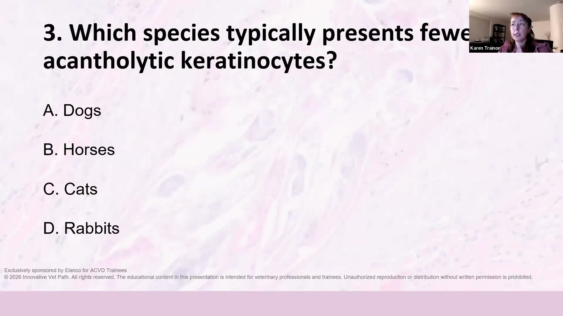 (A) Dogs (B) Horses (C) Cats ✅ (D) Rabbits
解析:貓對 acantholytic keratinocytes 很「小氣」;馬是第二名。→ 貓的 PF 切片找不到很多 acantholytic cells 時不要輕易排除。 → Part 1:1. Pemphigus foliaceus
Q4 ⏱ 54:12 — 狗出現 acantholytic keratinocytes 的三大原因?
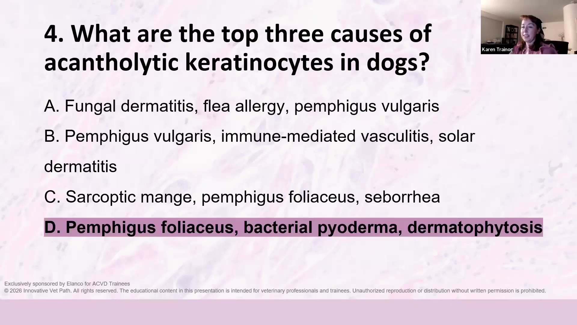 (A) Fungal dermatitis, flea allergy, pemphigus vulgaris (B) Pemphigus vulgaris, immune-mediated vasculitis, solar dermatitis (C) Sarcoptic mange, pemphigus foliaceus, seborrhea (D) Pemphigus foliaceus, bacterial pyoderma, dermatophytosis ✅
解析:PF、細菌性膿皮症、皮癬菌症 —— 她說「要熟到倒背如流」。(Part 1 另補第四個罕見原因:Darier’s-like disease) → Part 1:1. Pemphigus foliaceus
Q5 ⏱ 54:40 — 細胞學上,什麼線索支持 PF 而非細菌性膿皮症?
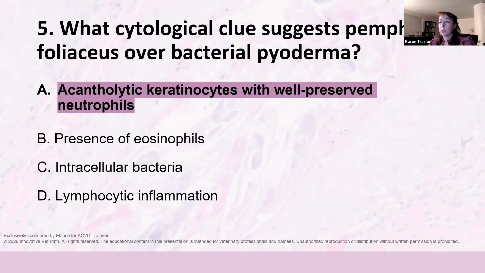 (A) Acantholytic keratinocytes with well-preserved neutrophils ✅ (B) Presence of eosinophils (C) Intracellular bacteria (D) Lymphocytic inflammation
解析:棘層鬆解細胞+保存良好(非退化性)的嗜中性球。若是細菌性膿皮症,會預期看到胞內細菌或退化性嗜中性球。 → Part 1:1. Pemphigus foliaceus
Q6 ⏱ 55:14 — Pemphigus vulgaris(PV,尋常型天疱瘡)的經典組織學發現?
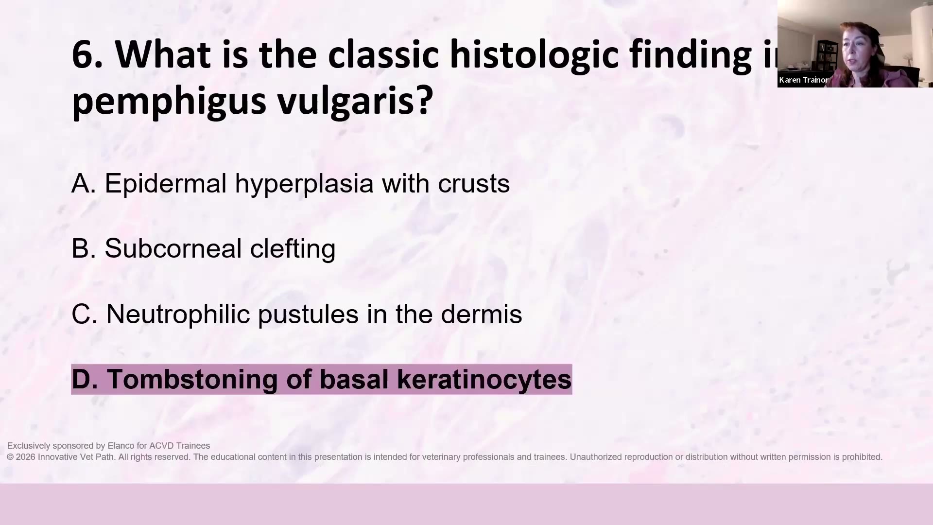 (A) Epidermal hyperplasia with crusts (B) Subcorneal clefting (C) Neutrophilic pustules in the dermis (D) Tombstoning of basal keratinocytes ✅
解析:基底角質細胞「墓碑狀」排列(tombstoning)—— 基底層上方裂開,基底細胞像一排墓碑留在地板上。 → Part 1:2. Pemphigus vulgaris
Q7 ⏱ 55:22 — PV 的主要自體抗原?
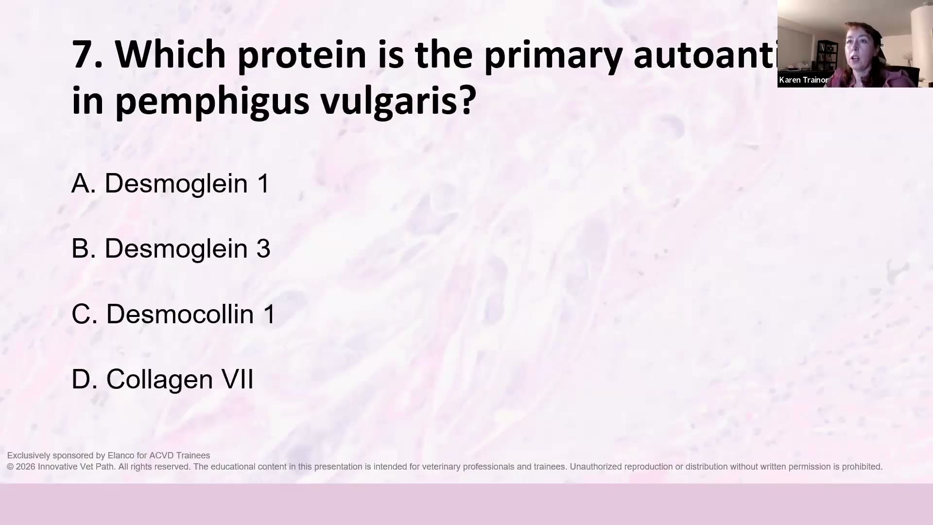 (A) Desmoglein 1 (B) Desmoglein 3 ✅ (C) Desmocollin 1 (D) Collagen VII
解析:Desmoglein 3。(對照:人類 PF 是 desmoglein 1、犬 PF 主要是 desmocollin 1;collagen VII 是 epidermolysis bullosa acquisita/dystrophic EB) → Part 1:2. Pemphigus vulgaris
Q8 ⏱ 55:54 — 確認 junctional epidermolysis bullosa(JEB,接合型表皮鬆解水疱症)用哪個特殊染色?
 (A) Ziehl-Neelsen (B) GMS (C) PAS (Periodic Acid-Schiff) ✅ (D) Masson’s trichrome
解析:PAS。ZN(Ziehl-Neelsen)是抗酸染色、GMS(Grocott’s methenamine silver)染黴菌、Masson’s trichrome 染膠原與 fibrin。 → Part 1:3. Junctional epidermolysis bullosa
Q9 ⏱ 56:18 — JEB 的裂隙發生在哪一層?
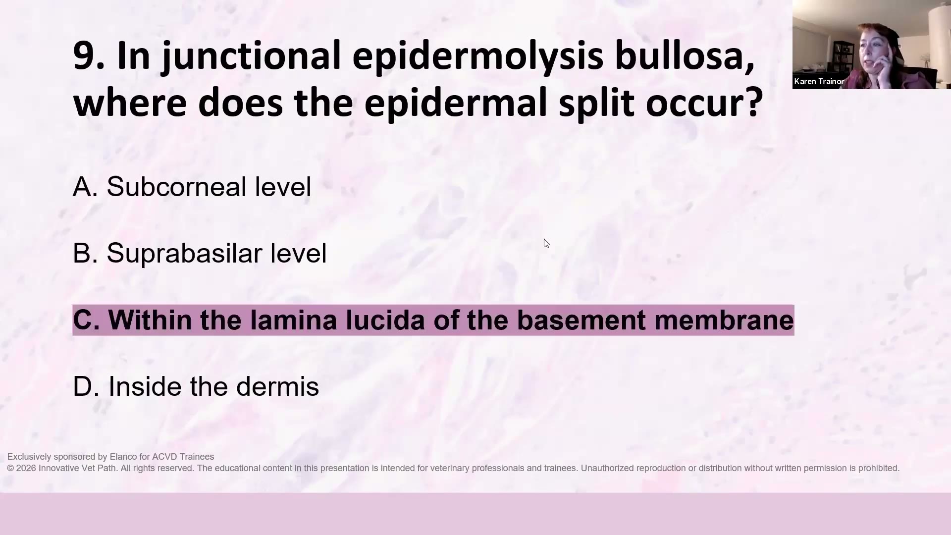 (A) Subcorneal level (B) Suprabasilar level (C) Within the lamina lucida of the basement membrane ✅ (D) Inside the dermis
解析:她說這是最刁鑽的一題:裂在基底膜的 lamina lucida(透明層)裡 → PAS 會把 lamina densa(緻密層)標在水疱的「地板」。 📖 她建議回去讀 Dr. Petra Bizikova 的犬貓水疱性疾病回顧文章(Part 1 的層次示意圖就是出自這篇,她說應該是 open access)。 → Part 1:3. Junctional epidermolysis bullosa
Q10 ⏱ 56:56 — 哪個犬種經典地與 exfoliative cutaneous lupus erythematosus(ECLE,剝落性皮膚紅斑性狼瘡)相關?
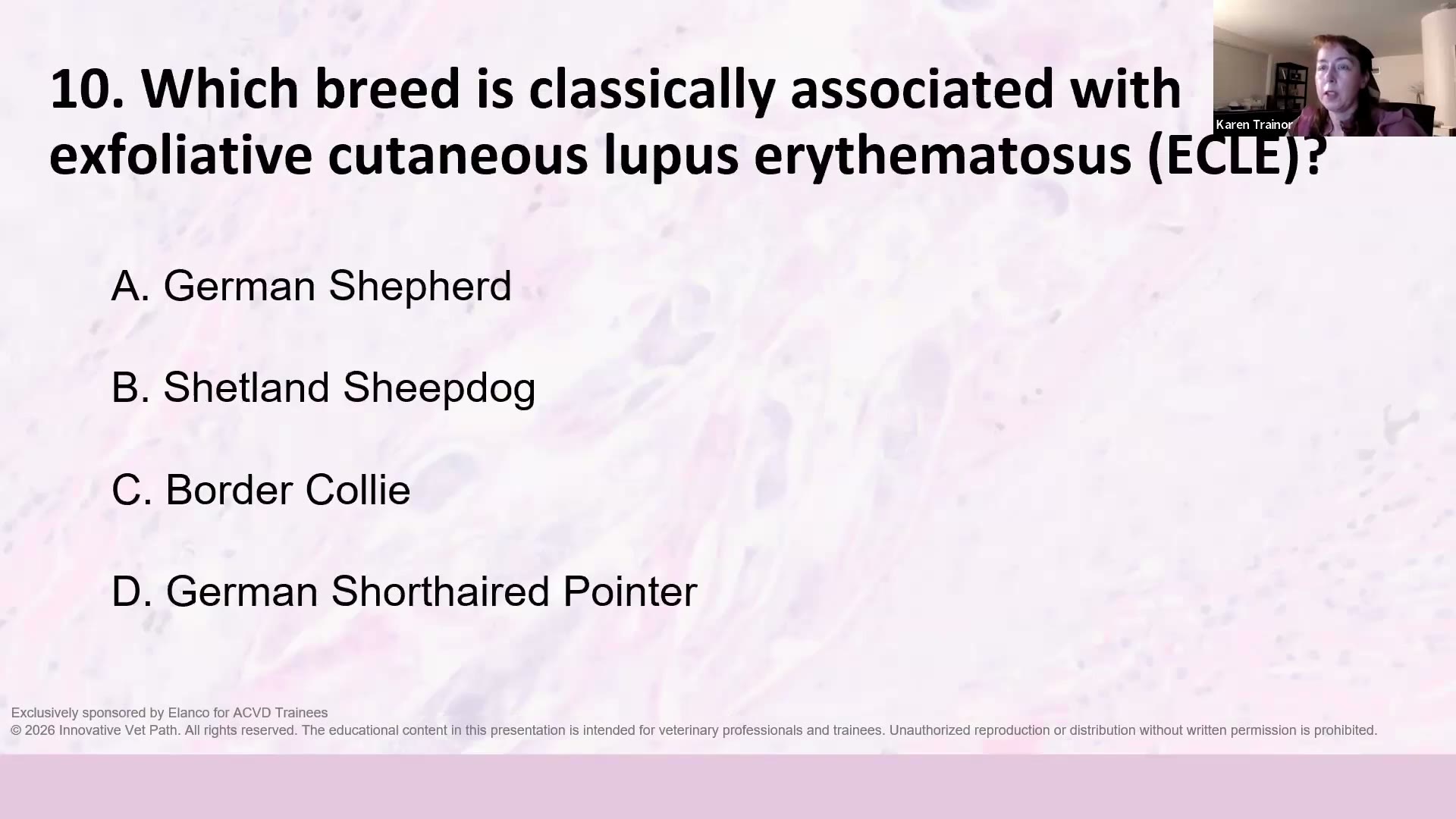 (A) German Shepherd (B) Shetland Sheepdog (C) Border Collie (D) German Shorthaired Pointer ✅
解析:德國短毛指示犬(GSHP)。陷阱:德國牧羊犬什麼病都會出現所以容易選錯;邊境牧羊犬與喜樂蒂是 vesicular cutaneous lupus(VCLE,水疱型皮膚紅斑性狼瘡)。 → Part 1:5. Exfoliative cutaneous lupus erythematosus of the GSHP
Q11 ⏱ 58:00 — 哪兩個組織學特徵合在一起對 ECLE 有特異性?
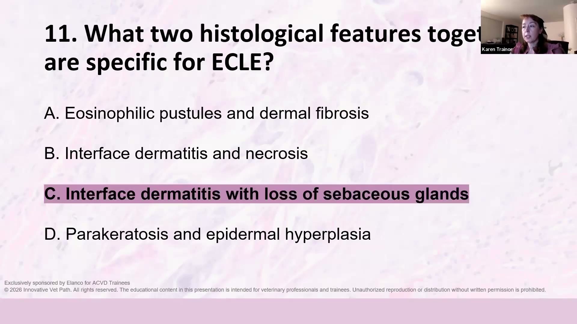 (A) Eosinophilic pustules and dermal fibrosis (B) Interface dermatitis and necrosis (C) Interface dermatitis with loss of sebaceous glands ✅ (D) Parakeratosis and epidermal hyperplasia
解析:Interface dermatitis +皮脂腺消失。皮脂腺掉光(sebaceous gland dropout)→ 表面才會有大量皮屑,所以叫「exfoliative」。她提醒這題是「很久以前講的,要把記憶撢一撢灰」。 → Part 1:5. Exfoliative cutaneous lupus erythematosus of the GSHP
Q12 ⏱ 58:14 — 哪個病的定義是 interface dermatitis + transepidermal apoptosis + lymphocyte satellitosis?
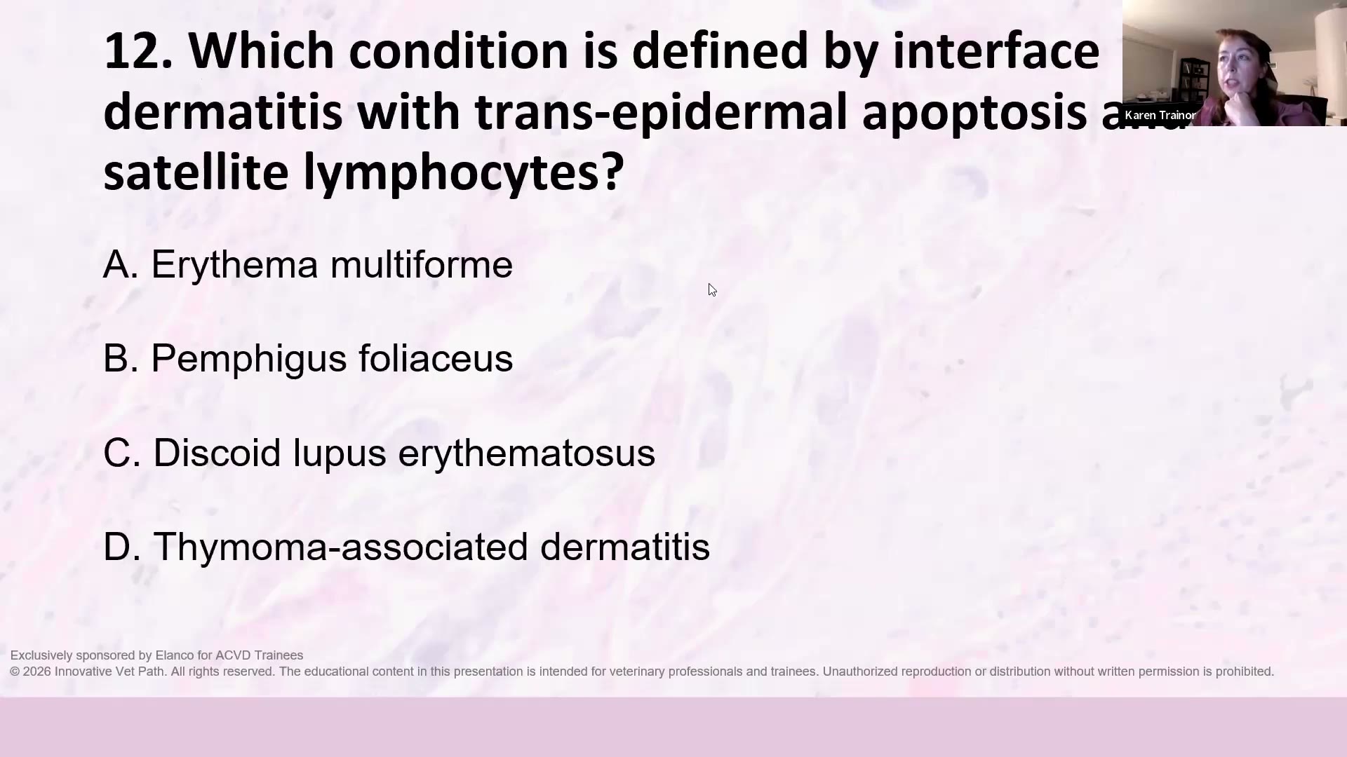 (A) Erythema multiforme ✅ (B) Pemphigus foliaceus (C) Discoid lupus erythematosus (D) Thymoma-associated dermatitis
解析:Erythema multiforme(EM,多形性紅斑)。 → Part 1:6. Erythema multiforme
Q13 ⏱ 59:02 — 組織學上什麼特徵能區分 EM 與 lupus?
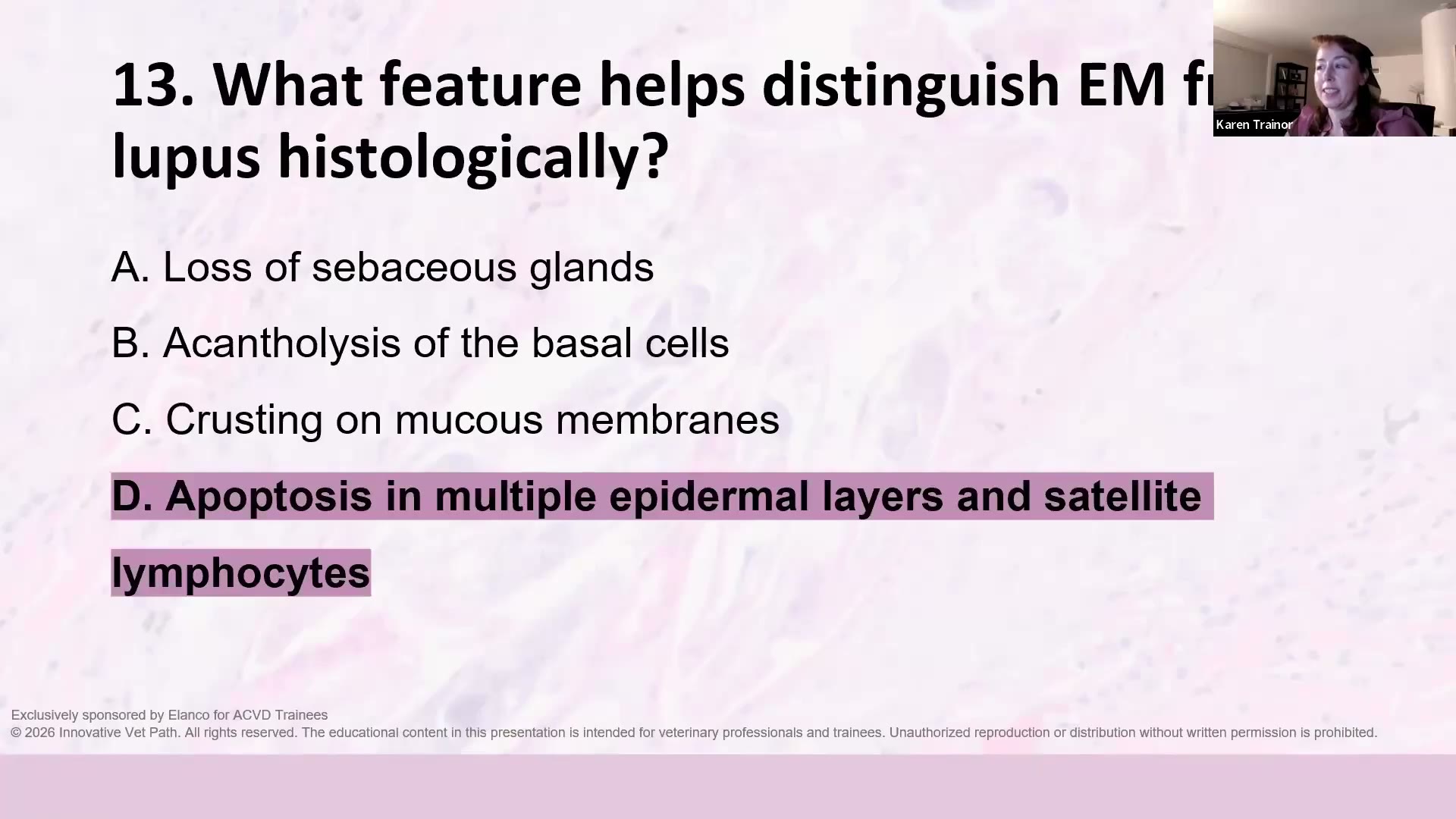 (A) Loss of sebaceous glands (B) Acantholysis of the basal cells (C) Crusting on mucous membranes (D) Apoptosis in multiple epidermal layers and satellite lymphocytes ✅
解析:表皮多層(全層)都有凋亡細胞+衛星淋巴球 → EM;lupus 的凋亡集中在基底層。 → Part 1:6. Erythema multiforme
Q14 ⏱ 59:36 — 貓 thymoma-associated exfoliative dermatitis(胸腺瘤相關剝落性皮膚炎) 的三個特徵?
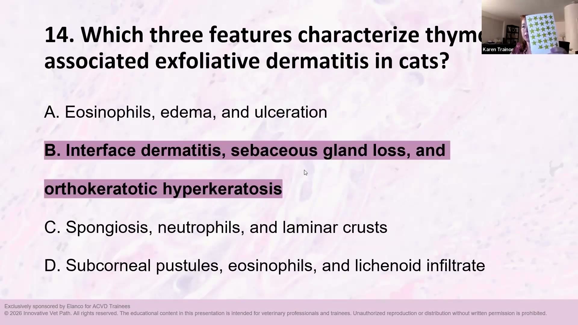 (A) Eosinophils, edema, and ulceration (B) Interface dermatitis, sebaceous gland loss, and orthokeratotic hyperkeratosis ✅ (C) Spongiosis, neutrophils, and laminar crusts (D) Subcorneal pustules, eosinophils, and lichenoid infiltrate
解析:Interface dermatitis +皮脂腺消失+ orthokeratotic hyperkeratosis。(字幕顯示她一開始口誤成「not thymoma associated」,隨即更正) → Part 1:8. (Thymoma-associated) Exfoliative dermatitis — feline
Q15 ⏱ 59:58 — 哪個病最常與 爪子脫落+甲床色素失禁(pigment incontinence) 相關?
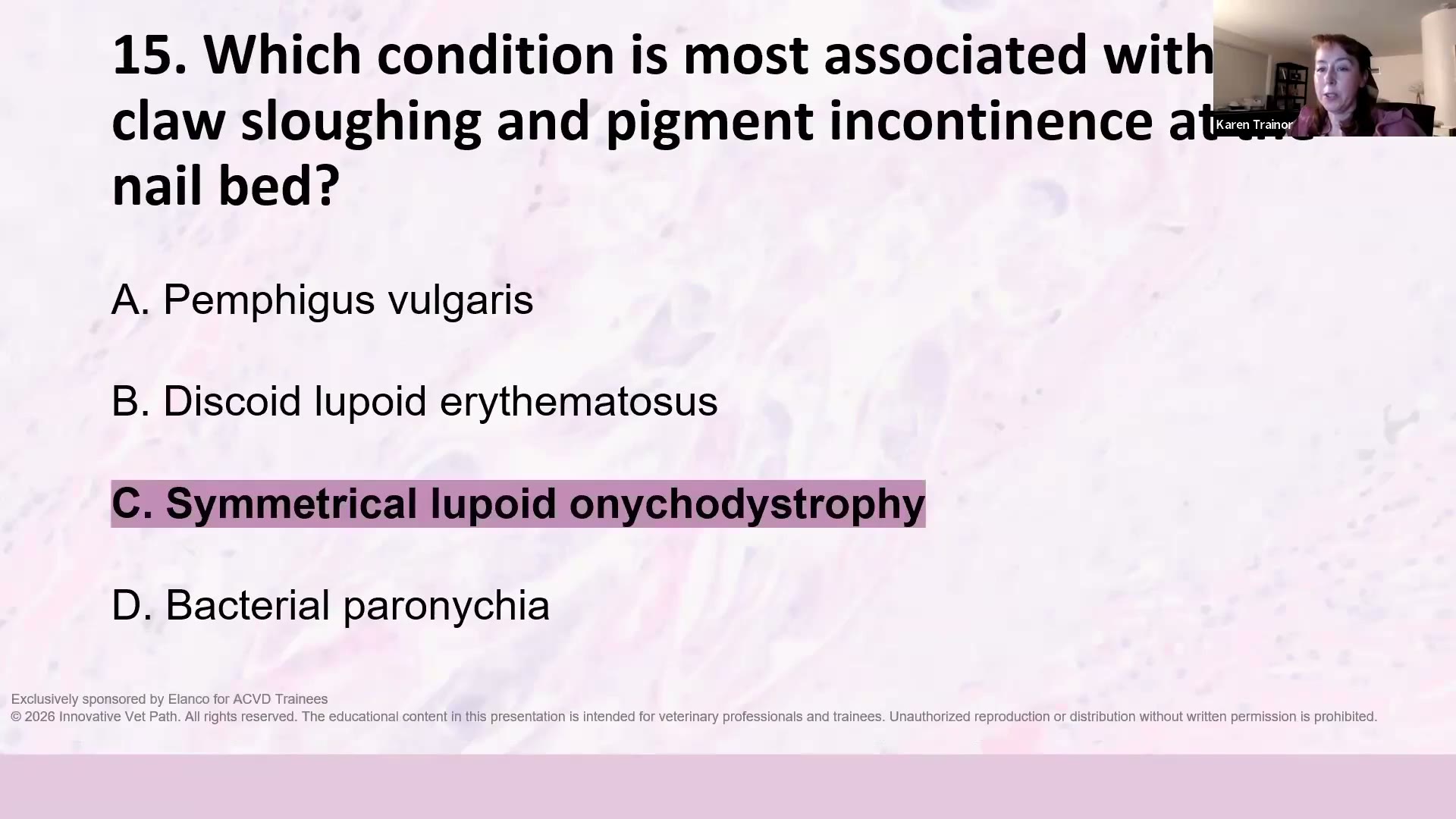 (A) Pemphigus vulgaris (B) Discoid lupoid erythematosus (C) Symmetrical lupoid onychodystrophy ✅ (D) Bacterial paronychia
解析:Symmetrical lupoid onychodystrophy(SLO,對稱性狼瘡樣爪甲失養症)。 → Part 1:9. Symmetrical lupoid onychodystrophy
Q16 ⏱ 60:18 — PAS 染色在 JEB 標示的是什麼?
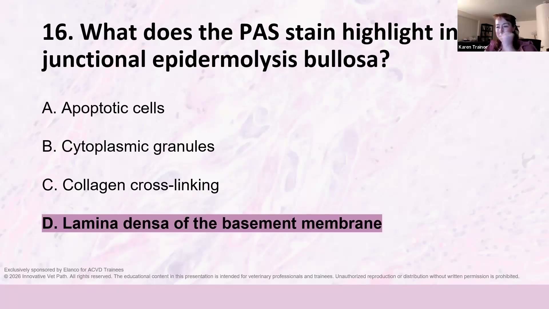 (A) Apoptotic cells (B) Cytoplasmic granules (C) Collagen cross-linking (D) Lamina densa of the basement membrane ✅
解析:基底膜的 lamina densa —— 與第 9 題是一組:裂在 lamina lucida,PAS 陽性的 lamina densa 留在水疱地板。(字幕漏錄這題,答案取自投影片揭曉畫面) → Part 1:3. Junctional epidermolysis bullosa
Q17 ⏱ 60:42 — 為什麼診斷 discoid lupus erythematosus(DLE,盤狀紅斑性狼瘡)時,臨床醫師與病理醫師的溝通很重要?
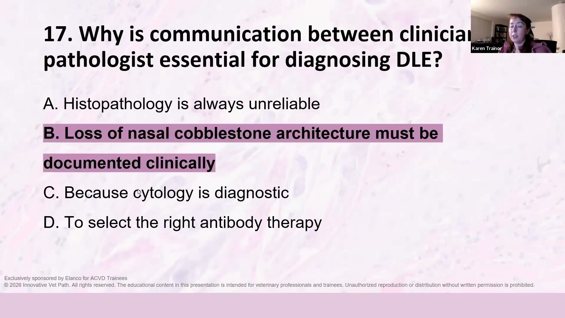 (A) Histopathology is always unreliable (B) Loss of nasal cobblestone architecture must be documented clinically ✅ (C) Because cytology is diagnostic (D) To select the right antibody therapy
解析:鼻鏡鵝卵石紋理消失必須由臨床記錄。可以附照片、寫在病史描述,兩者都給最好。「如果病理醫師只拿到切片、完全不知道臨床外觀,對誰都不好玩。」 → Part 1:10. Discoid lupus erythematosus
Q18 ⏱ 61:32 — Hepatocutaneous syndrome(HCS,肝皮症候群)的經典組織學 pattern?
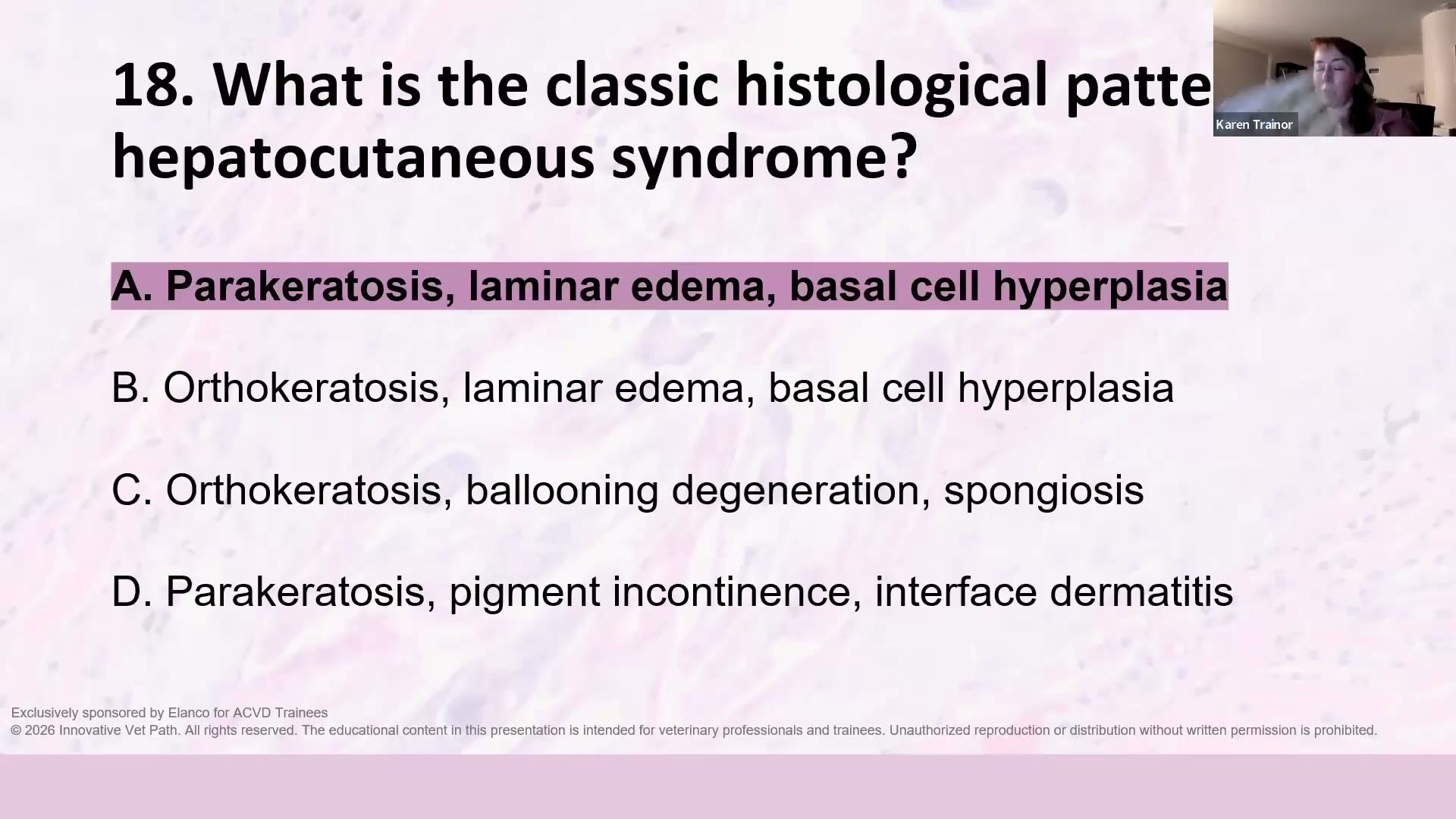 (A) Parakeratosis, laminar edema, basal cell hyperplasia ✅ (B) Orthokeratosis, laminar edema, basal cell hyperplasia (C) Orthokeratosis, ballooning degeneration, spongiosis (D) Parakeratosis, pigment incontinence, interface dermatitis
解析:Red(parakeratosis)— White(laminar edema)— Blue(basal cell hyperplasia)。她說「mock exam 不可能沒有這題」。陷阱是 B:把 para- 換成 ortho-。 → Part 1:11. Hepatocutaneous syndrome
Q19 ⏱ 61:36 — Split paw pad disease(腳掌墊裂開症)的裂隙在哪裡?
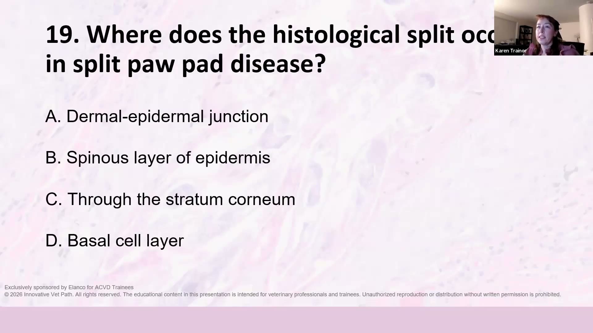 (A) Dermal-epidermal junction (B) Spinous layer of epidermis (C) Through the stratum corneum ✅ (D) Basal cell layer
解析:穿過 stratum corneum(角質層) —— 不是全層裂開。 → Part 1:12. Split paw pad disease
Q20 ⏱ 62:16 — 貓 eosinophilic plaque(嗜伊紅性斑塊)的特徵性組織學表現?
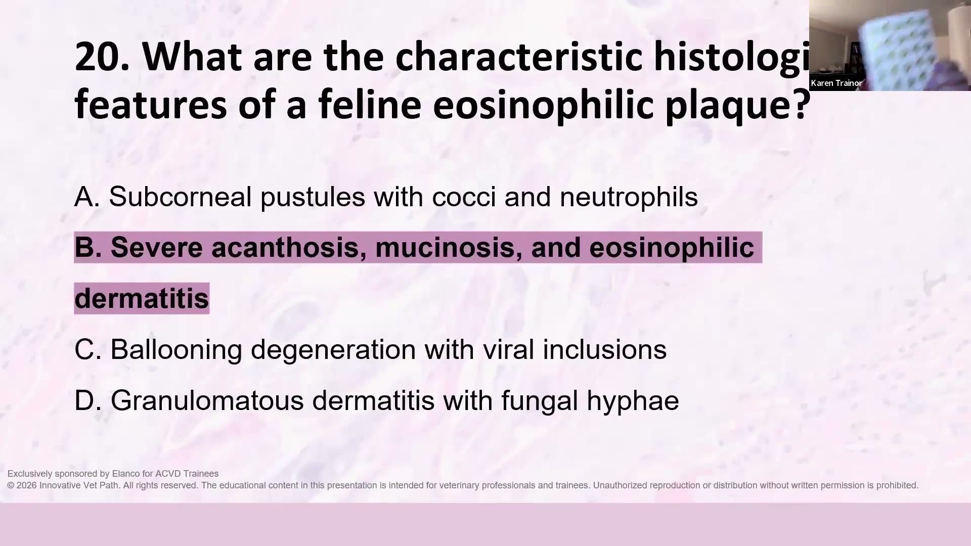 (A) Subcorneal pustules with cocci and neutrophils (B) Severe acanthosis, mucinosis, and eosinophilic dermatitis ✅ (C) Ballooning degeneration with viral inclusions (D) Granulomatous dermatitis with fungal hyphae
解析:嚴重 acanthosis + mucinosis +嗜伊紅球性皮膚炎。 → Part 1:14. Feline eosinophilic plaque
✍️ 自我測驗表(遮住上面答案重做一次)
| # | 考點 | 正解 | 我的答案 | 對錯 |
|---|---|---|---|---|
| 1 | Pemphigus foliaceus | B | ||
| 2 | Pemphigus foliaceus | B | ||
| 3 | Pemphigus foliaceus | C | ||
| 4 | Pemphigus foliaceus | D | ||
| 5 | Pemphigus foliaceus | A | ||
| 6 | Pemphigus vulgaris | D | ||
| 7 | Pemphigus vulgaris | B | ||
| 8 | Junctional epidermolysis bullosa | C | ||
| 9 | Junctional epidermolysis bullosa | C | ||
| 10 | Exfoliative cutaneous lupus erythematosus of the GSHP | D | ||
| 11 | Exfoliative cutaneous lupus erythematosus of the GSHP | C | ||
| 12 | Erythema multiforme | A | ||
| 13 | Erythema multiforme | D | ||
| 14 | (Thymoma-associated) Exfoliative dermatitis — feline | B | ||
| 15 | Symmetrical lupoid onychodystrophy | C | ||
| 16 | Junctional epidermolysis bullosa | D | ||
| 17 | Discoid lupus erythematosus | B | ||
| 18 | Hepatocutaneous syndrome | A | ||
| 19 | Split paw pad disease | C | ||
| 20 | Feline eosinophilic plaque | B |
📝 我的問題(這場沒被回答 → 寄信問)
Trainor 說:有想補充的疾病或問題,可以先 email 給她(karen@innovativevetpath.com),她會在這場回答或另外做教材。
- Part 1 提到 Darier’s-like disease 可能有新文獻/NAVDF 摘要 → 目前進度?診斷標準?
- Split paw pad disease 的 KRT5 假說她持保留態度 → 目前證據到哪裡?
- Symmetrical lupoid onychodystrophy 在貓 的病例是否已投稿/可否分享特徵?
- Plasma cell pododermatitis 與腎臟病變的 2026 JFMS 新論文,完整書目是?
- Alopecia areata 那篇「Morgan 的論文」完整書目?
✅ 我的行動清單
- 把 7 個腫瘤的 buzzword 做成 Anki → 專科考試記憶宮殿
- 遮住答案重做 20 題 mock exam,答錯的回 Part 1 重讀
- 找 Petra Bizikova 的犬貓水疱性疾病回顧文章(🚨 完整書目待查),建 🔴 資訊源筆記
- 把上面的問題整理成一封信寄給 karen@innovativevetpath.com
- 10 月 DermPath 101 報名資訊留意 email
- 門診實作:老貓單一趾頭腫 → 趾頭+胸腔 X 光;馬的腫塊採檢避開潰瘍、要有完整表皮
🔑 關鍵字
- Karen Trainor Board Prep 2026 總覽
- 2026.08.22 Board Review Part 1
- 2026.08.28 Board Review Part 2
- ECVD HistoRound 考題庫(依主題分類)
- 專科考試記憶宮殿
- DermPath