2026.08.06 ECVD HistoRound(自動整理)
由
螢幕錄影 2026-08-06 晚上11.00.32.mov(70:36,21 GB)自動產生:whisper.cpp large-v3-turbo 逐字稿(英文原文,未翻譯)+ ffmpeg 定點切片截圖。 ⚠️ 這是機器自動整理的原始版,逐字稿可能有小錯。已濃縮版本見 2026.08.06 ECVD HistoRound。 🔊 平均音量 −27.4 dB(有旁白)。講師:Chiara Brachelente、Ilaria Porcellato。 ✂️ 實際討論到 01:03:00 結束;之後只有掛線的環境音,逐字稿出現「bye / thank you」重複是 whisper 對靜音段的幻聽,已保留但請忽略。
- 已人工校對/濃縮 → 2026.08.06 ECVD HistoRound
🖼️ 切片畫面(定點擷取,對應各案例教學重點)
[00:01:05]
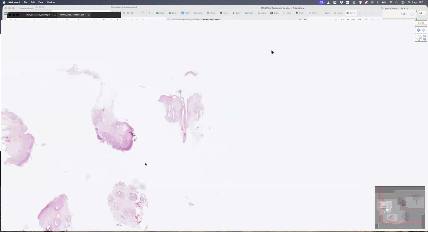
[00:03:20]
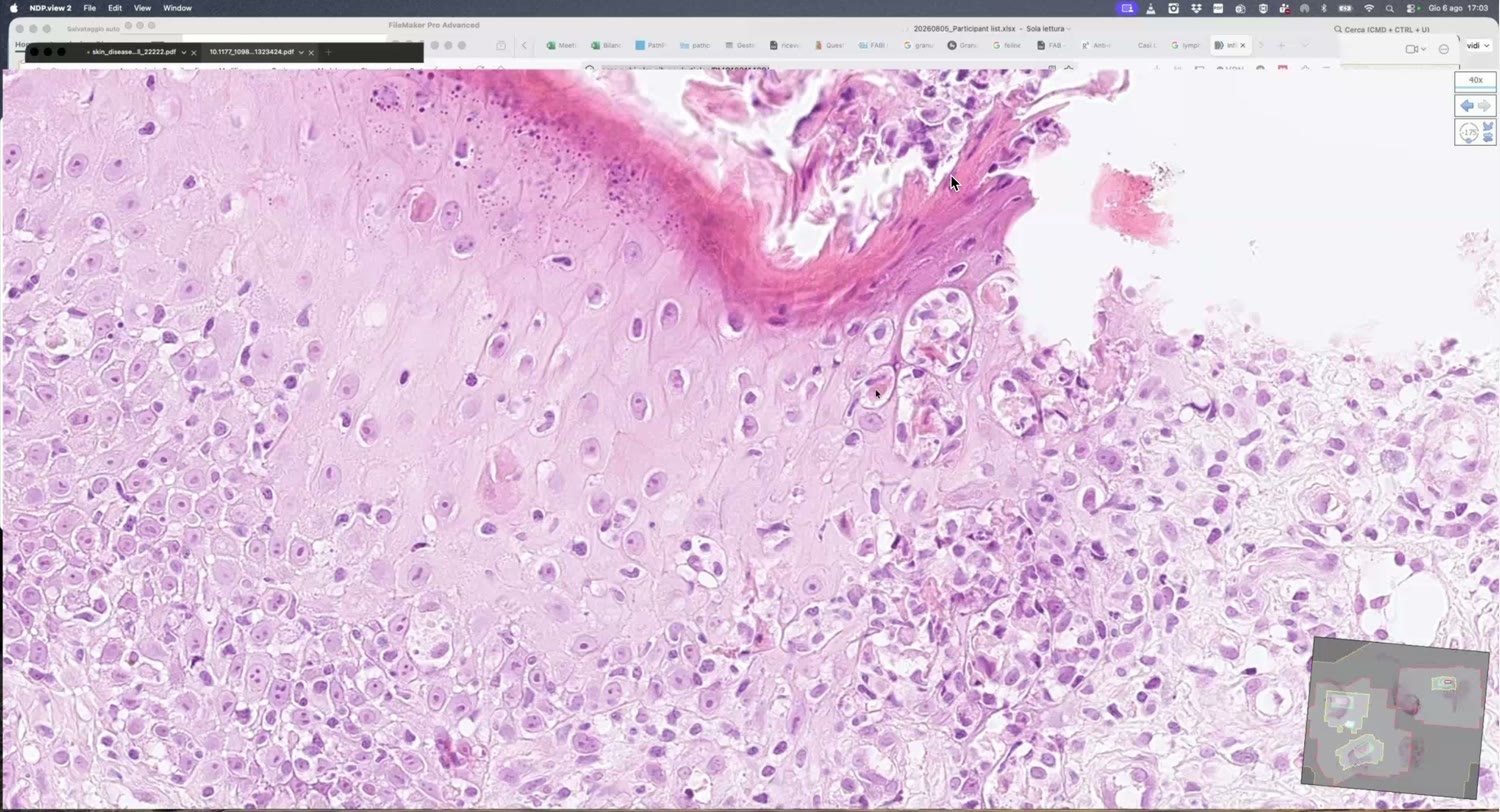
[00:07:45]
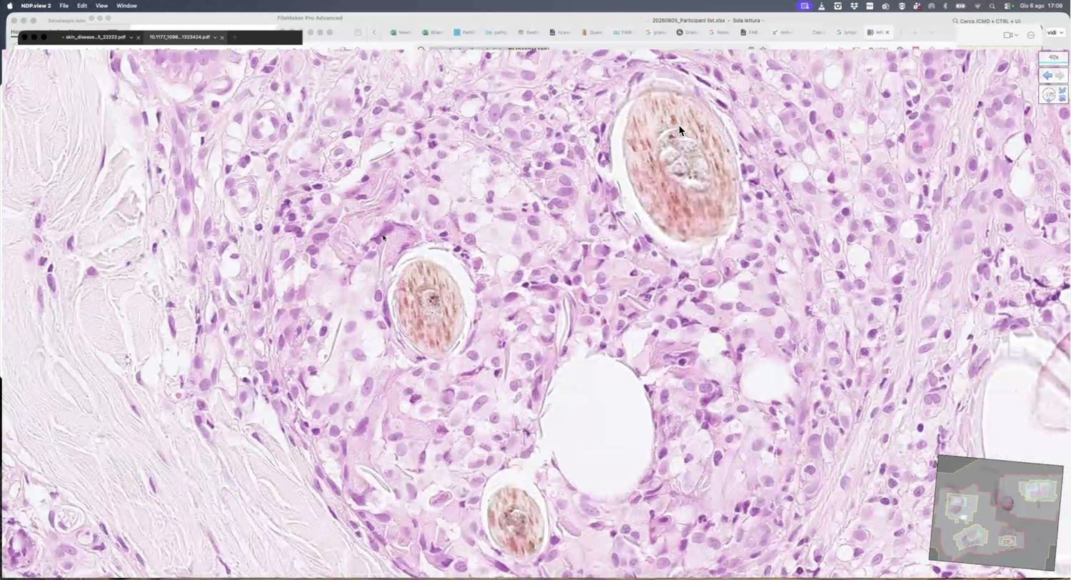
[00:08:45]
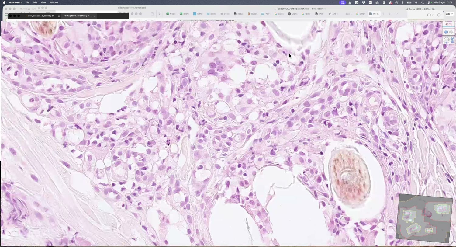
[00:12:45]
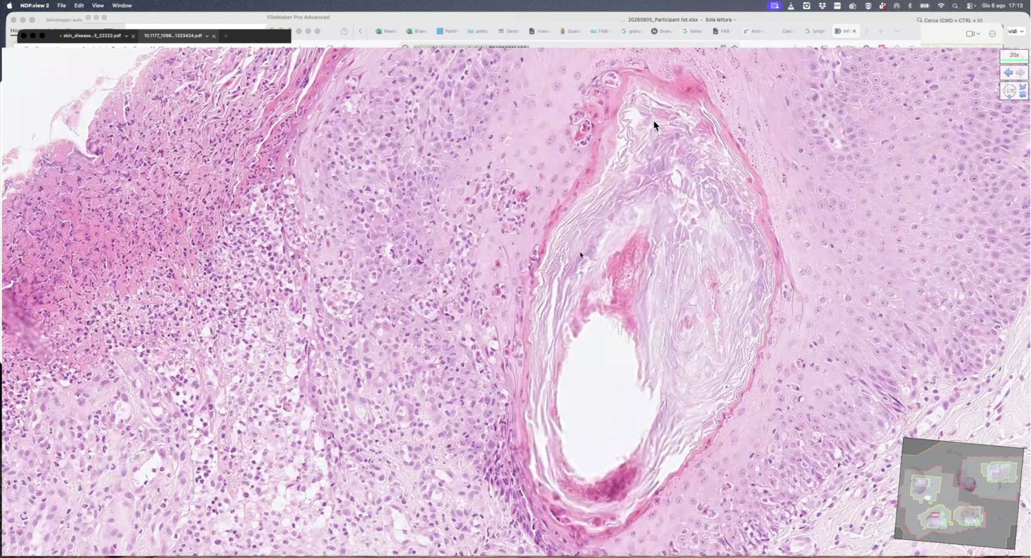
[00:23:10]
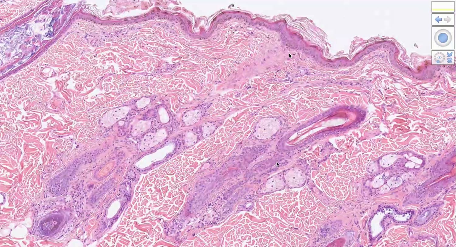
[00:27:50]
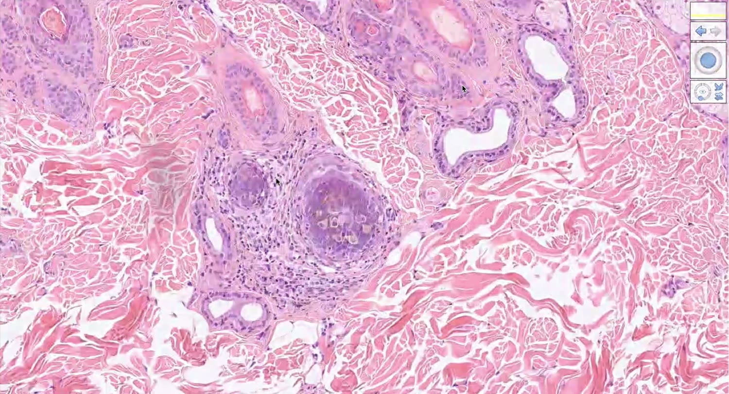
[00:28:50]
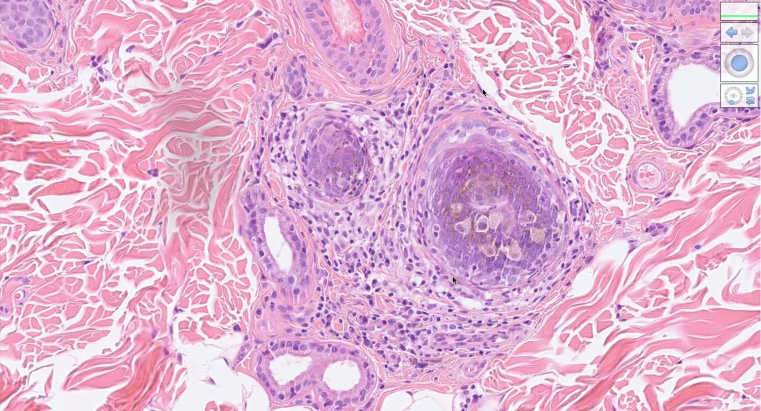
[00:29:25]
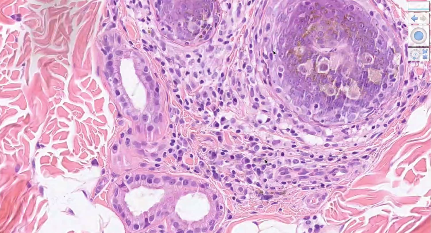
[00:30:05]
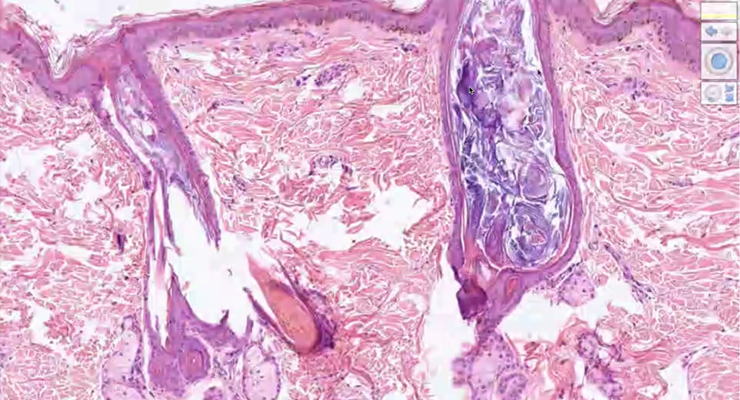
[00:36:10]
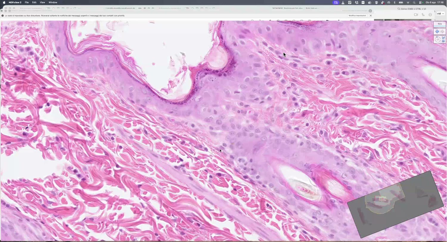
[00:37:20]
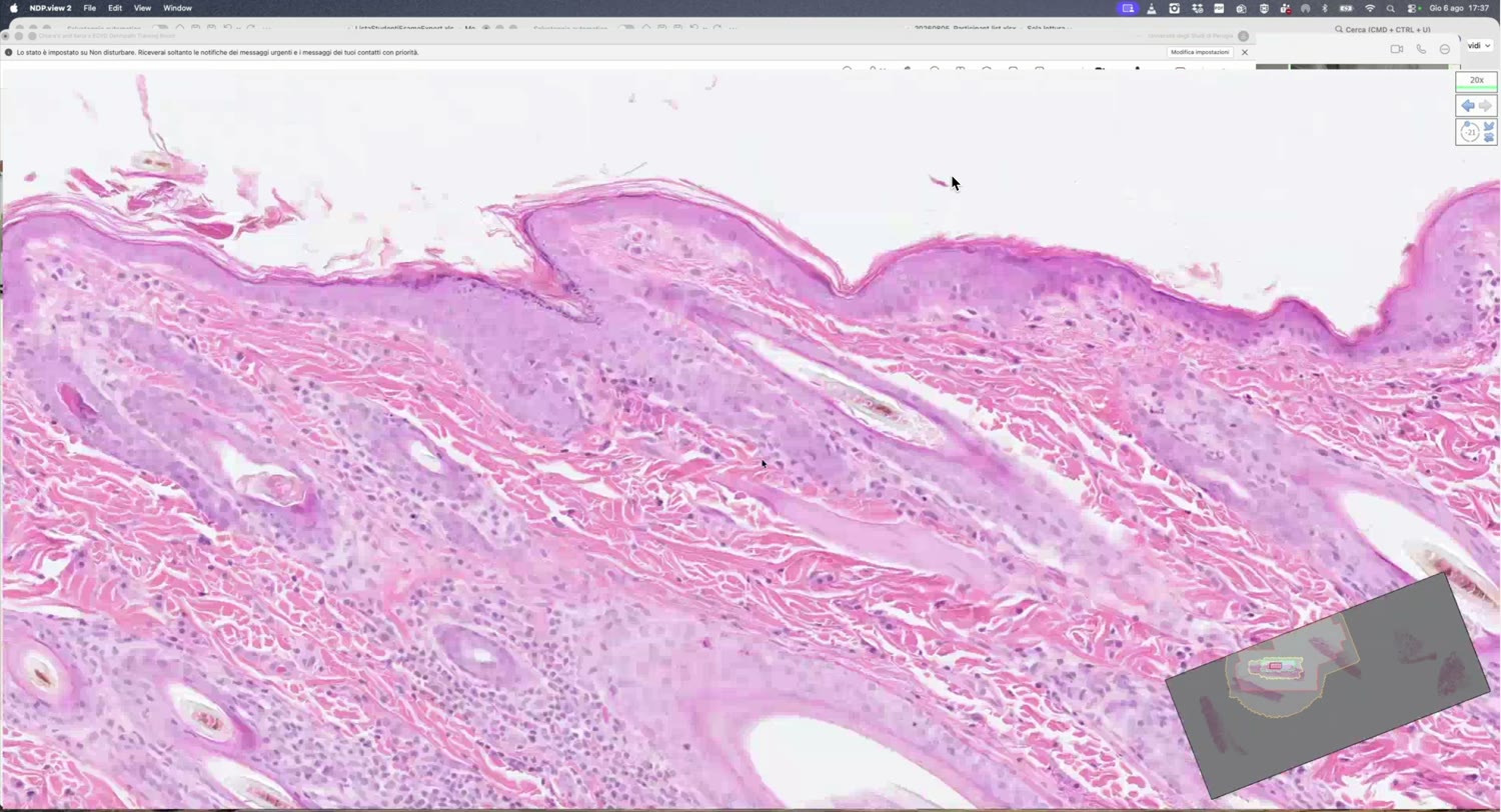
[00:41:00]
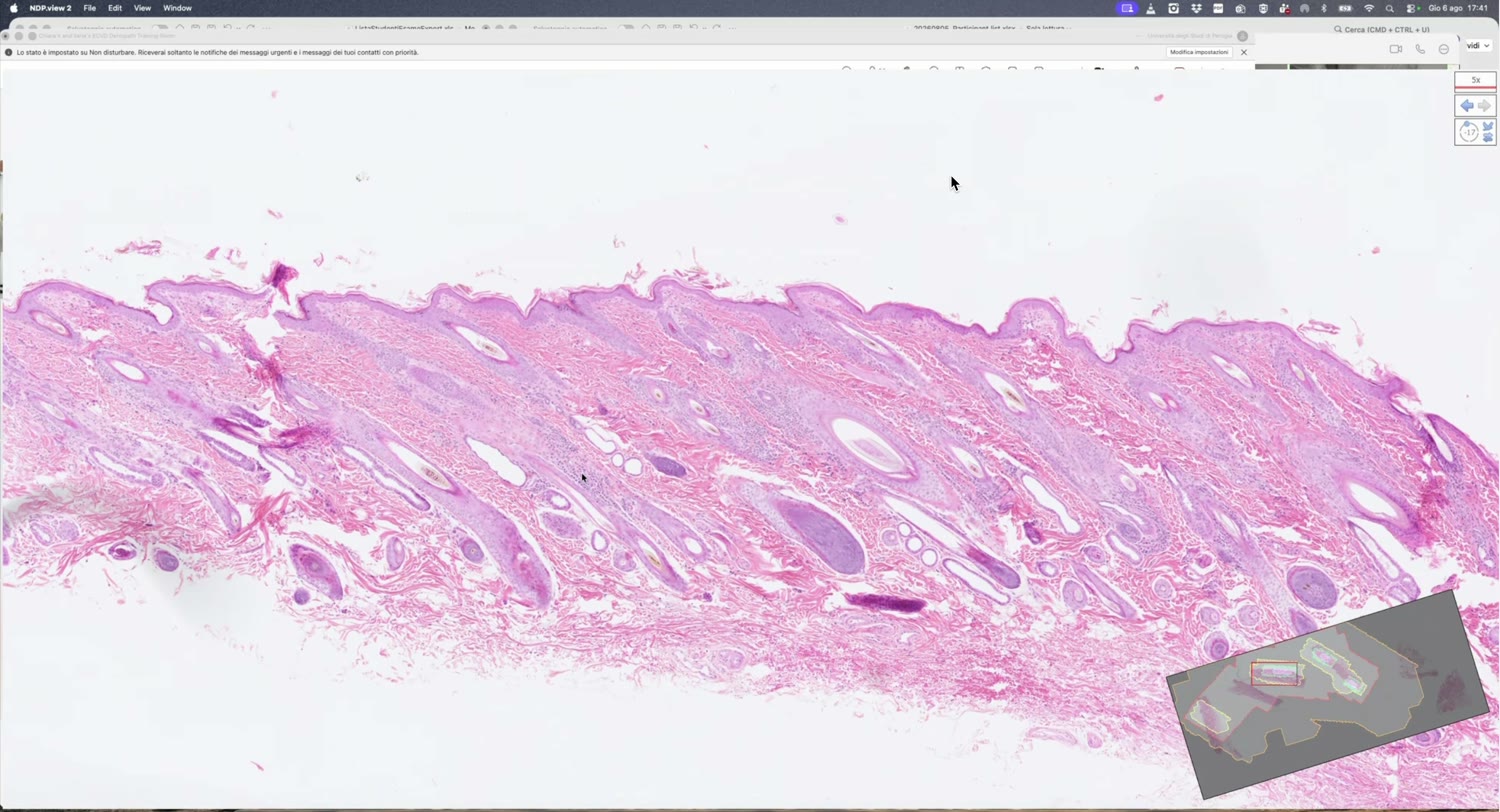
[00:42:20]
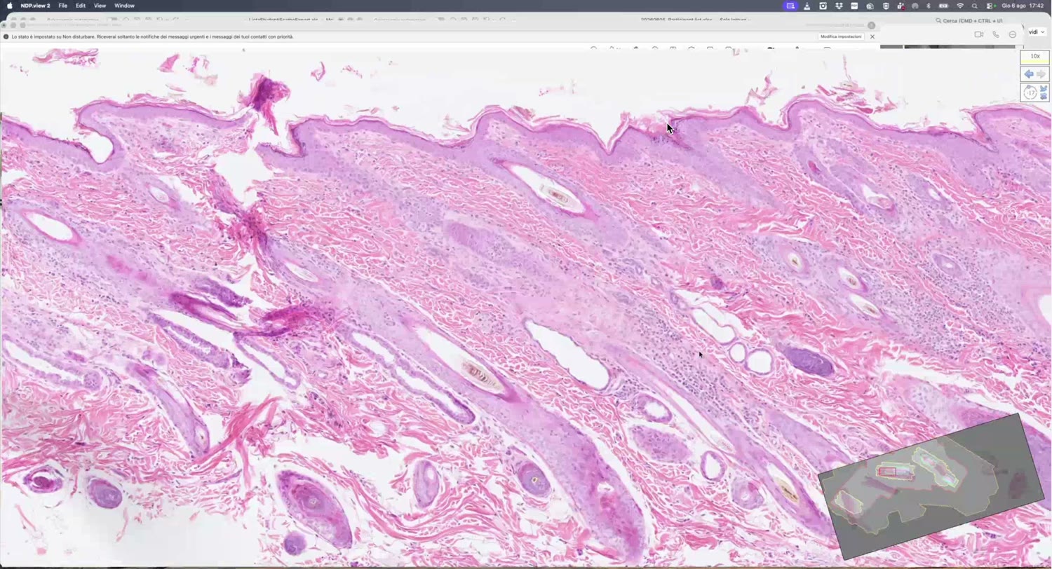
[00:45:45]
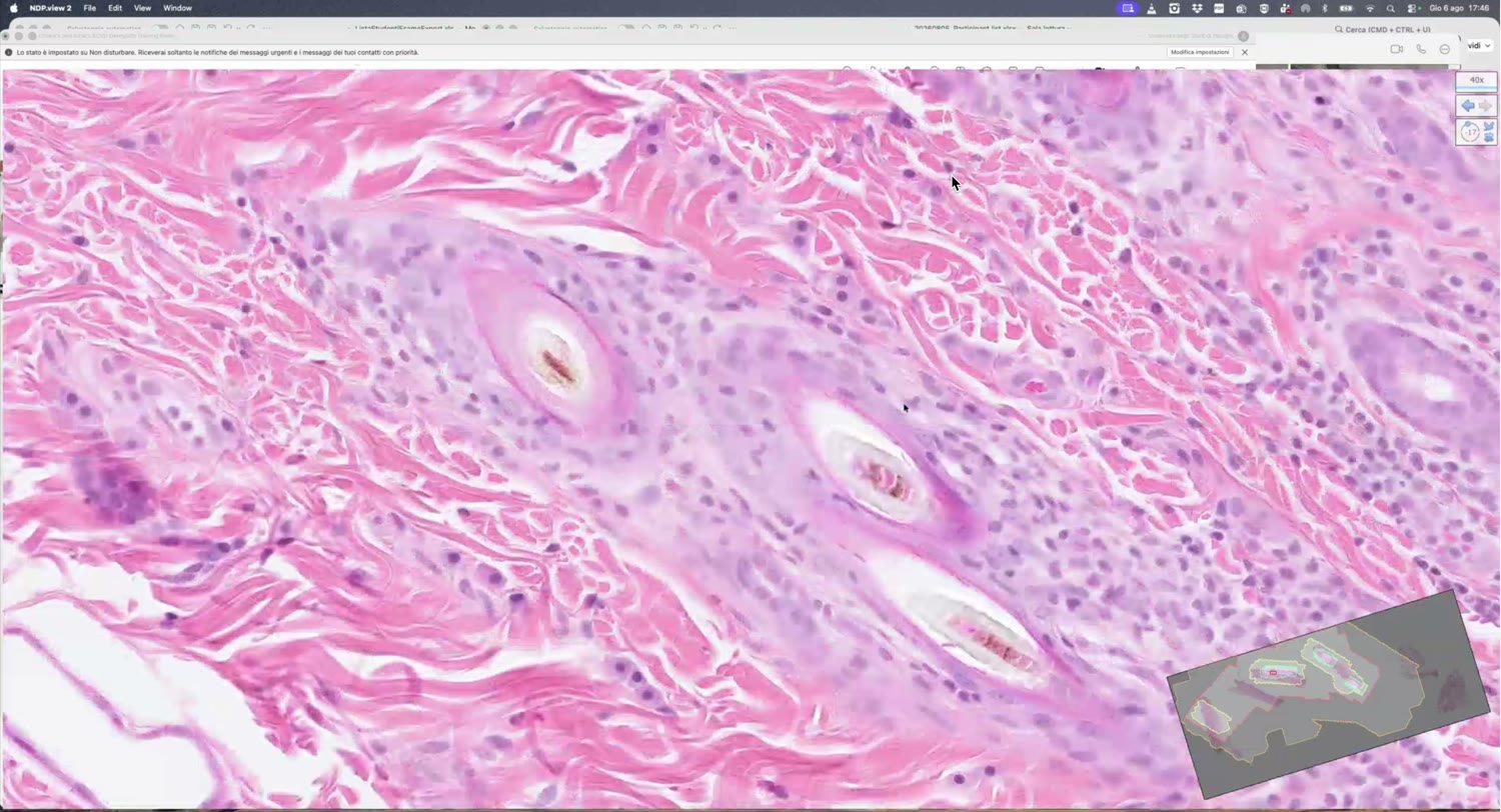
[00:46:30]
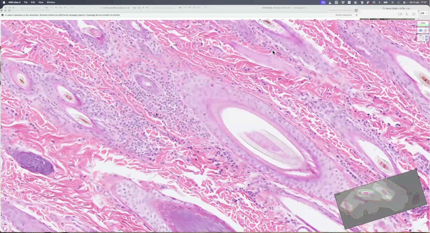
[00:48:20]
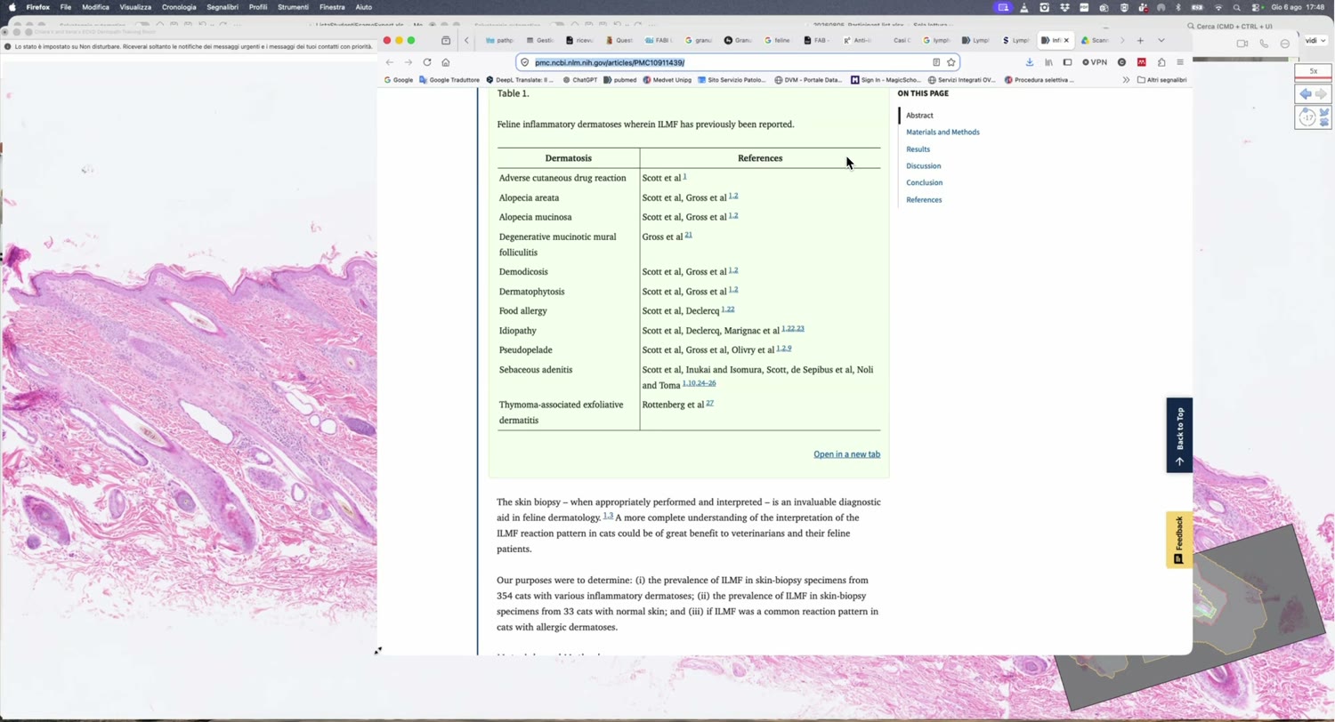
[00:59:55]
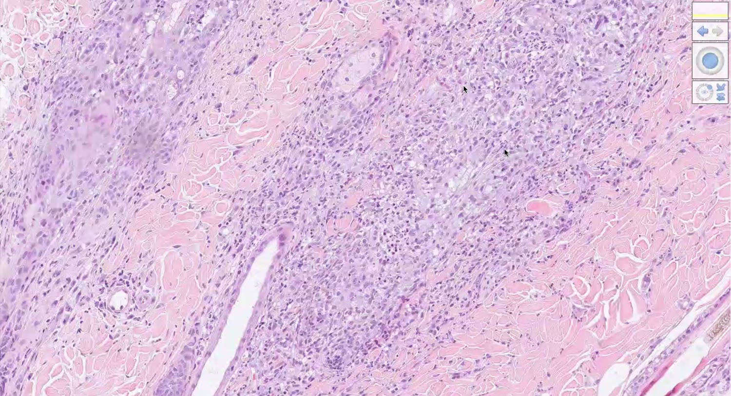
[01:00:30]
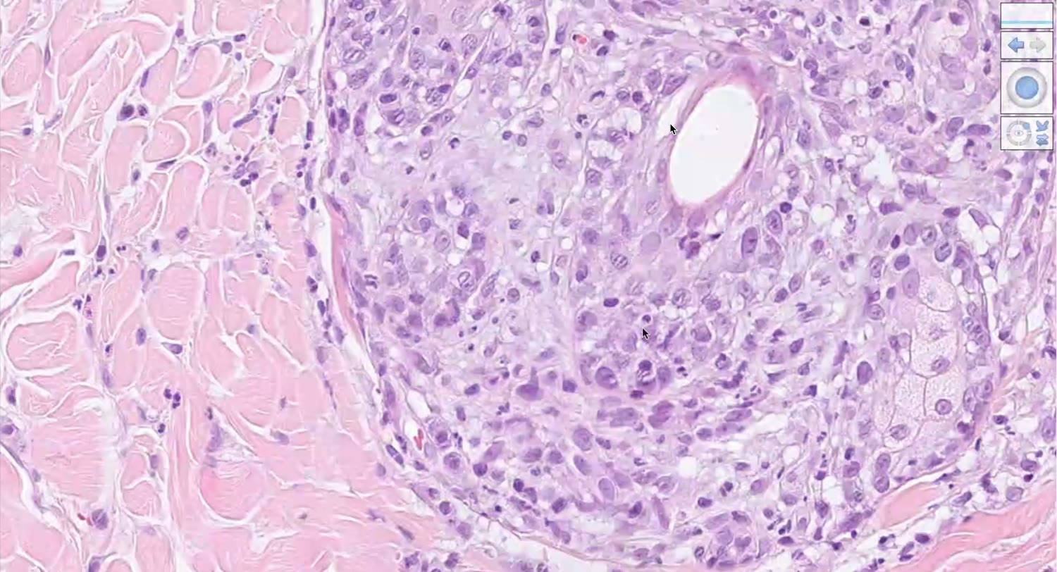
[01:01:25]
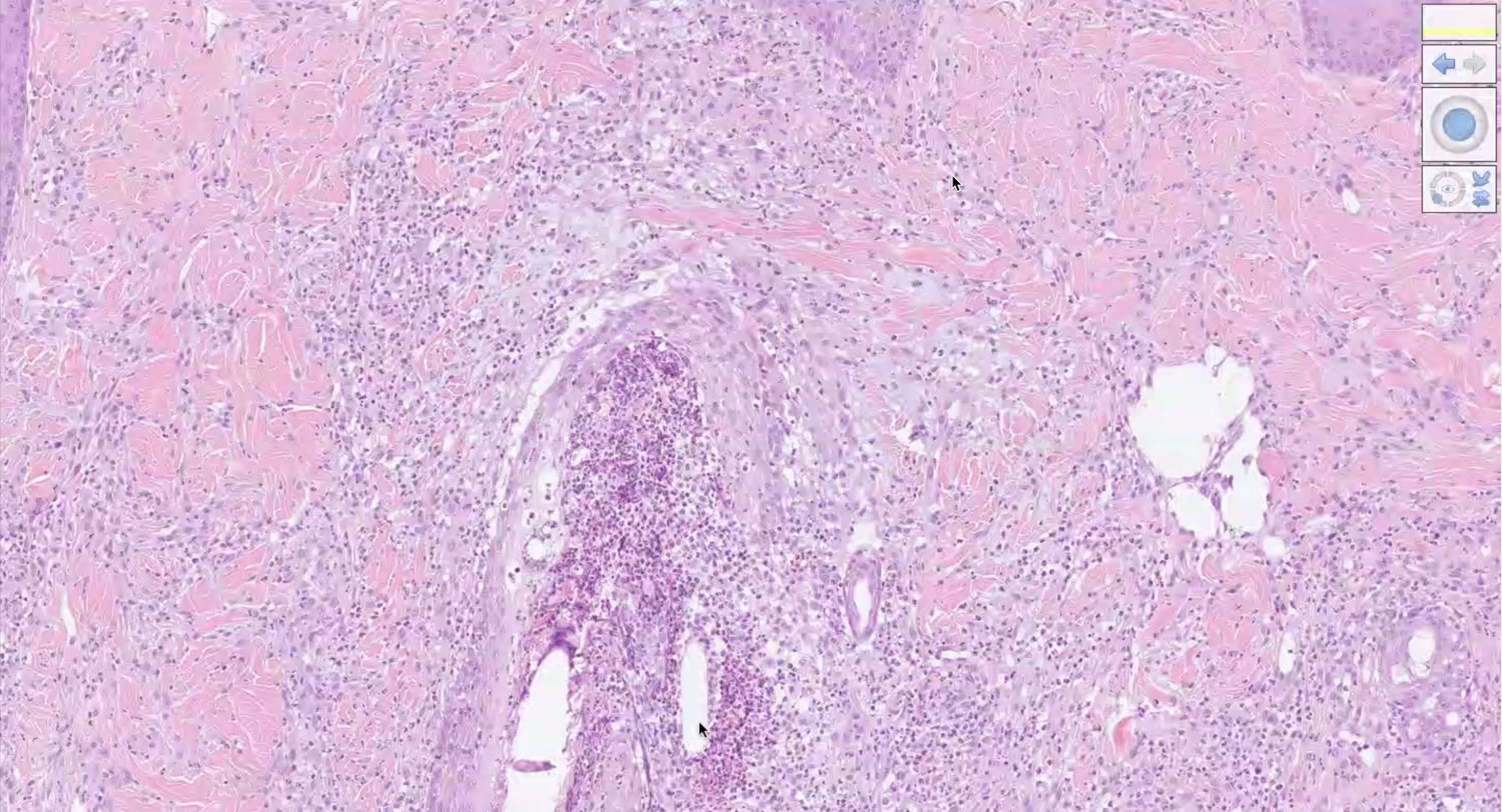
[01:01:40]
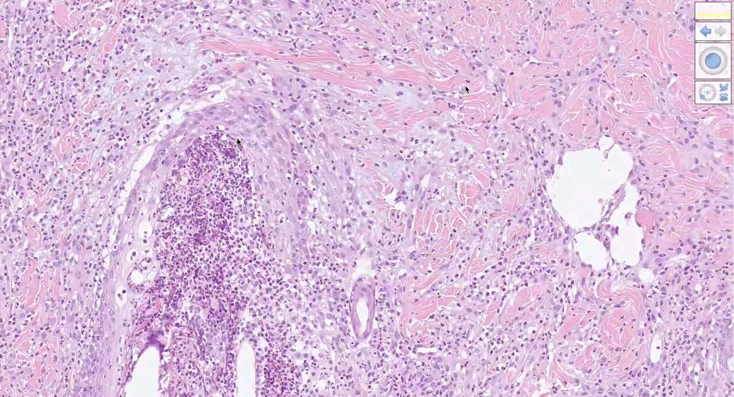
📝 逐字稿(英文原文)
[00:00:00] So I think we can start and I will start with the first slide. [00:00:10] So if someone wants to describe the slide is B599. [00:00:23] Do you see my screen? [00:00:24] Yes, that’s mine and I will try to describe it, but I’ve had quite a hard time [00:00:29] with that one, to be honest. [00:00:31] So Mia, right? [00:00:33] Okay. [00:00:34] Yes. [00:00:35] Please go ahead. [00:00:36] So there are several cuts with a lot of, I think it’s necrotic tissue as well, at least [00:00:47] in the epidermis. [00:00:50] So it was actually quite hard to find a place where there was no necrotic tissue, but I think [00:00:55] there were two pieces where you could see parts of the epidermis, at least. [00:01:00] Yes, exactly. [00:01:01] This one. [00:01:02] And then I think the lower left one as well. [00:01:06] Yeah. [00:01:07] Yeah. [00:01:08] That’s the one. [00:01:09] So in general, it seemed to be a pretty diffuse, superficial pattern and also, I think, perifollicular [00:01:21] pattern. [00:01:22] Mm-hmm. [00:01:23] Yes. [00:01:24] And when going in, the epidermis was, with most of the parts, not really recognizable [00:01:33] anymore. [00:01:34] Mm-hmm. [00:01:35] But how do you call this? [00:01:40] Interface? [00:01:41] No, I mean, when you say that the epidermis is not recognizable anymore and it’s missing, [00:01:51] what are you describing? [00:01:56] It’s just an answer, right? [00:01:58] Oh, okay. [00:01:59] Yes. [00:02:00] Yeah. [00:02:01] Sorry. [00:02:02] It’s just, yeah. [00:02:03] Okay. [00:02:04] Mm-hmm. [00:02:05] Yes. [00:02:06] So yeah, there were like several ulcerations and also necrotic epidermis. [00:02:11] Yes. [00:02:12] And with the parts where the epidermis was still intact, it seemed to be hypoplastic. [00:02:20] Mm-hmm. [00:02:21] Yeah. [00:02:22] Wait a second. [00:02:23] Where was it? [00:02:25] Here? [00:02:26] Yes. [00:02:27] Yes. [00:02:28] Mm-hmm. [00:02:29] Yeah. [00:02:30] So yeah, I think that was most of the stuff that I wrote down for the epidermis. [00:02:37] And what is all of these things here? [00:02:42] So there is, I think, spongiosis in that part of the epidermis. [00:02:47] Yeah. [00:02:48] And there were also several apoptotic keratinocytes that you could see. [00:02:53] Yes. [00:02:54] And how do you describe these cells here? [00:02:58] These phenomenon here, where you have lymphocytes? [00:03:04] Satellitosis? [00:03:05] The satellitosis, you should have lymphocytes surrounding… [00:03:10] Oh, no. [00:03:11] Never mind. [00:03:12] Yeah. [00:03:13] Yes. [00:03:14] No, it’s okay. [00:03:15] Because it’s like here, for example. [00:03:17] Yes. [00:03:18] You see? [00:03:19] There is an apoptotic keratinocyte that is surrounded by lymphocytes. [00:03:24] But these cells, these lymphocytes that are going through the epidermis… [00:03:29] Exocytosis. [00:03:30] It’s called lymphocytic exocytosis. [00:03:32] Yes. [00:03:33] Right. [00:03:34] Okay. [00:03:35] Mm-hmm. [00:03:36] Yes. [00:03:38] And so, I mean, if we go closer to the areas where the epidermis is going into necrosis and [00:03:53] it’s alterating, we can see that there are these areas of many apoptotic keratinocytes that [00:04:01] are surrounded by lymphocytes. [00:04:05] So there is satellitosis. [00:04:07] And we also can imagine that all of these are rests of apoptotic keratinocytes. [00:04:14] Right? [00:04:15] Mm-hmm. [00:04:16] Okay. [00:04:17] Mm-hmm. [00:04:18] Okay. [00:04:19] Yes. [00:04:20] And then I was trying to look more into the cell infiltrate itself. [00:04:33] Mm-hmm. [00:04:34] Which one? [00:04:36] Which infiltrate are you talking about? [00:04:39] The superficial dermal cell infiltrate. [00:04:43] Yeah. [00:04:44] Yeah. [00:04:45] Which was… [00:04:46] Actually, I thought it’s like a bunch of eosinophils, but it doesn’t look like being eosinophils [00:04:56] anymore here. [00:04:57] Mm-hmm. [00:04:58] I did… [00:04:59] For sure, lymphocytes. [00:05:00] Yes. [00:05:01] But the lymphocytes are these, huh? [00:05:05] Okay? [00:05:06] Mm-hmm. [00:05:07] You see the error, right? [00:05:09] Mm-hmm. [00:05:10] Mm-hmm. [00:05:11] So the majority of the cells are not lymphocytes. [00:05:15] What are these big cells with elongated and sometimes like bean-shaped nuclei with open chromatin? [00:05:28] Not as dark as the chromatin of lymphocytes. [00:05:33] Sorry. [00:05:34] Sorry. [00:05:35] These are… [00:05:36] They’re all… [00:05:37] Sorry. [00:05:38] I mean, I think they’re all like macrophages. [00:05:41] Yes. [00:05:42] Exactly. [00:05:43] And if you look at them, they are like 70% macrophages and 30% lymphocytes. [00:05:50] Mm-hmm. [00:05:51] Yes. [00:05:52] Okay. [00:05:57] And then I was also looking at the perifollicular area. [00:06:03] Mm-hmm. [00:06:04] Yes. [00:06:07] I could barely see any intact sebaceous glands. [00:06:12] Yeah. [00:06:13] I mean, they are somehow affected by the inflammation. [00:06:21] You can recognize them here, for example. [00:06:24] Mm-hmm. [00:06:25] Okay. [00:06:26] Mm-hmm. [00:06:27] I mean, to me, more than the sebaceous glands, other… [00:06:32] The follicular… [00:06:33] Yeah. [00:06:34] …area. [00:06:35] Yeah. [00:06:36] Yeah. [00:06:37] You see, like here, for example, the follicular wall is completely gone. [00:06:40] Yeah. [00:06:41] We just have the ghosts of the hair shafts and the hair follicle wall is just remaining in [00:06:50] some areas, but in other areas is completely affected. [00:06:55] Okay. [00:06:56] And you really see that the… [00:07:00] How do you say? [00:07:01] That the contour of what was the hair follicle wall has been replaced by these macrophagic and [00:07:12] lymphocytic infiltration. [00:07:13] Mm-hmm. [00:07:14] Yeah. [00:07:15] Yeah. [00:07:16] Okay. [00:07:17] And I just, I don’t know if you… [00:07:18] I will go in this area here because in this area we don’t have the epidermis and we just [00:07:33] have the next cell structure that are cut transversally. [00:07:39] And I mean, as a demonstration of the macrophagic nature, you also see multinucleated giant cells. [00:07:48] Mm-hmm. [00:07:49] Right? [00:07:50] That are eating up. [00:07:51] What are all these things here? [00:07:55] Mm-hmm. [00:07:56] You see this structure that I’m pointing at? [00:08:01] Mm-hmm. [00:08:02] Or not? [00:08:03] Okay. [00:08:04] Yes, I do. [00:08:05] These are just keratin lamelle. [00:08:07] Ah, okay. [00:08:08] Okay. [00:08:09] So, it’s the same thing that you see surrounding the hair shaft, right? [00:08:14] So, it’s really a granulomatous inflammation that is centered around the follicles and it’s [00:08:23] facing the hair follicle wall. [00:08:26] Yeah. [00:08:27] Do you agree? [00:08:28] Yeah. [00:08:29] I mean, it’s pretty obvious with that. [00:08:33] Okay. [00:08:34] With that. [00:08:35] Yeah. [00:08:36] And some, I mean, there are not very many, but there are a couple of multinucleated giant [00:08:43] cells. [00:08:44] And there are also a few neutrophils. [00:08:49] See? [00:08:50] Oh, yes. [00:08:51] Yeah. [00:08:52] Here, here it’s one. [00:08:55] And there is another one here. [00:08:58] Another one here. [00:08:59] So, it’s a, in this case, I would say granulomatous. [00:09:04] But when the, if the neutrophils are a bit more, I would call it probably piogranulomatous. [00:09:16] Okay. [00:09:17] Do you, did you notice anything else? [00:09:22] I probably should have, but I did not. [00:09:28] No, no, no, no, no, no. [00:09:30] It was just like, like you mentioned, for example, that this vicious glands. [00:09:35] Yeah. [00:09:36] That’s the only thing that I did notice that there were most of, I mean, I saw, I think, [00:09:40] two or three spots where they looked somewhat normal. [00:09:43] Yeah. [00:09:44] But most of them were kind of destroyed or look destroyed. [00:09:49] Yeah. [00:09:50] See here. [00:09:51] This is a nice rest of the vicious cells surrounded also by this inflammation. [00:09:59] Okay. [00:10:00] Okay. [00:10:01] So I agree with you that there is a partial involvement of the, partial involvement of the sebaceous glands. [00:10:09] Whereas for example, the apocrine glands, they look totally fine. [00:10:14] Okay. [00:10:15] Yep. [00:10:16] And what else? [00:10:17] I think there were scattered apoptotic cells, even in the wall of the hair follicles. [00:10:26] Mm-hmm. [00:10:27] So basically the same, or at least partially the same findings that we discussed in the, in the epidermis. [00:10:40] Yep. [00:10:41] And one can really see that the follicle is starting to be kind of attacked by the inflammation. [00:10:53] And then it kind of explodes or, I mean, it, it is destroyed by the inflammation. [00:11:00] Okay. [00:11:01] Yeah. [00:11:02] Um, this is another area and you still can see the apoptotic cells with the satellitosis. [00:11:13] And the apoptotic cells are at which level of the epidermis are they? [00:11:19] They are predominantly in the, like, they are basal and in the. [00:11:34] You see, but this is the basal layer and this is the superficial layer. [00:11:40] So basically. [00:11:41] Epidermal basically. [00:11:43] Yeah. [00:11:44] And, uh, so whenever you have these, uh, basically we have this, uh, dichotomy. [00:11:51] So either they are basal or suprabasal, or they are panepidermal, transepidermal. [00:11:59] Like in this case, because in this case you have apoptotic cells at all layers of the epidermis. [00:12:06] Okay. [00:12:07] So, and this pattern here is called the cytotoxic dermatitis because the, um, cytotoxicity basically [00:12:19] is, uh, caused by these, uh, T lymphocytes that are most likely CD8 cytotoxic T lymphocytes [00:12:28] that, um, induce, uh, um, keratinocytes apoptosis. [00:12:33] Okay. [00:12:34] Okay. [00:12:35] And this is another nice example of, uh, or, uh, more apoptotic cells. [00:12:43] Um, here we have apoptotic cells in the hair follicle. [00:12:48] And the ones that we looked at before they were in the epidermis. [00:12:53] So it’s both, uh, epidermal and follicular cytotoxicity. [00:12:58] Okay. [00:12:59] Okay. [00:13:00] Okay. [00:13:01] And then probably, uh, in the description, you should make some cleanup because, uh, if you [00:13:10] describe the necrosis and the ulceration, uh, then you should, uh, here, for example, there [00:13:20] inflammation just below, uh, the epidermis and are these, uh, um, infiltrating neutral fields [00:13:28] that are secondary to the ulceration. [00:13:31] And then there is probably edema, vessel congestion, never forget all these, uh, clean up because, [00:13:43] uh, at the exam, it gives you points. [00:13:46] Okay. [00:13:47] Mm-hmm. [00:13:48] So never. [00:13:49] Is that then basically kind of the morph or is that a step before the morph? [00:13:54] No, no. [00:13:55] This is the, what is called the cleanup. [00:13:58] Uh, so you, you have described the main findings that are the, um, I don’t anticipate it because [00:14:09] you have to put it in the morph, but everything we have described in terms of what is happening [00:14:15] to the follicles and what is happening to the epidermis is the main finding of the, of [00:14:21] the lesion. [00:14:22] And then the, I would say that the, the cleanup is the, um, uh, necrosis, the alteration, the, [00:14:31] uh, granulation tissue, uh, proliferation with inflammatory cells and so on. [00:14:36] Okay. [00:14:37] For example, if you have bacteria in the crust, it’s, it’s a secondary finding, right? [00:14:43] It’s secondary to the ulceration. [00:14:46] Yep. [00:14:47] Right. [00:14:48] Right. [00:14:49] Okay. [00:14:50] So do we wanna, um, uh, one thing that probably is, uh, um, important to say, um, let’s try [00:15:01] to make the, um, morphologic diagnosis. [00:15:04] Okay. [00:15:05] Or, uh, I can ask you, what are for you the most important findings in this case? [00:15:15] What do you think are the primary findings and what are the secondary findings? [00:15:19] So some of them we have already discussed, but what do you think is the main, um, target here? [00:15:30] I think the main, um, I mean, the main things are the necrosis, the ulceration, the epidermal [00:15:43] epidermal and follicular involvement. [00:15:45] Mm-hmm. [00:15:46] Um, the cytotoxic, um, unepidermal pattern. [00:15:56] Mm-hmm. [00:15:57] Um, and the, probably the macrophage and lymphocyte predominant. [00:16:01] Pattern? [00:16:02] Mm-hmm. [00:16:03] Or inflammation? [00:16:04] Do you all, as you are first ear, do you all, um, approach the lesion in terms of trying [00:16:13] to recognize the basic is a pathologic pattern? [00:16:17] Yes or no? [00:16:18] I don’t know. [00:16:20] Because you have, you have several patterns. [00:16:26] What I’m talking about is a very vascular dermatitis, cytotoxic interface, postular, folliculitis, [00:16:39] perifolliculitis, funclosis, paniculitis, vasculitis. [00:16:41] Okay. [00:16:42] So if you try to put this slide in one of these boxes, one or two, one or two, okay. [00:16:53] Only. [00:16:54] Do you explain all the lesions with only cytotoxic dermatitis, panepidermal? [00:17:02] I don’t explain the follicular lesions with it. [00:17:07] Yeah. [00:17:08] Yeah. [00:17:09] What would be the morphologic diagnosis for the follicles? [00:17:14] Moral folliculitis? [00:17:16] Mm-hmm. [00:17:17] What, what can you add to the Moral folliculitis? [00:17:22] Do you know that with the Moral folliculitis, you have to go a step further? [00:17:27] Because there are different types of Moral folliculitis. [00:17:29] There is the Moral Pustular, Moral Infiltrative, Moral Isinophilic, Moral. [00:17:37] How would be in this case here? [00:17:45] Um. [00:17:46] If we have described this guy here. [00:17:50] It’s with the multi-nucleated giant cells. [00:17:56] Mm-hmm. [00:17:57] Then what would be the Moral? [00:17:58] If you see a multi-nucleated giant cell. [00:18:01] If you see macrophages. [00:18:03] If you see… [00:18:04] Granulomatous? [00:18:05] Yeah, exactly. [00:18:06] So it’s a granulomatous Moral folliculitis. [00:18:10] Yeah. [00:18:11] In this case, the two main patterns here are the granulomatous Moral folliculitis and the cytotoxic [00:18:19] panepidermal dermatitis. [00:18:20] Mm-hmm. [00:18:21] Okay? [00:18:22] Yeah. [00:18:23] The alteration, for example, if you look at the patterns, there is no ulcerative pattern. [00:18:36] Mm-hmm. [00:18:37] Because the alteration, both clinically and histologically, is always a secondary lesion. [00:18:44] Mm-hmm. [00:18:45] Okay? [00:18:46] It can be due to a vasculitis. [00:18:48] It can be due to single-cell apoptosis, such in this case. [00:18:53] It can be due to scratching. [00:18:55] It can be due to thermal burn. [00:18:58] But it’s never purely alteration. [00:19:04] You know what I mean? [00:19:05] Yeah. [00:19:06] Okay. [00:19:07] So what would you think is the cause here? [00:19:14] Or the mechanism, let’s say, more than the cause. [00:19:19] If you have a cytotoxic panepidermal dermatitis, what would your differential diagnosis be? [00:19:31] I mean, all of the cytotoxic diseases. [00:19:35] Mm-hmm. [00:19:36] So starting from lupus to EM to… [00:19:39] But lupus is not panepidermal. [00:19:42] Yes. [00:19:43] So it would be more… [00:19:44] Lupus is interface. [00:19:45] Yes. [00:19:46] So it would be more EM or Steven Johnson’s or TEN. [00:19:50] Exactly. [00:19:51] So what would be the mechanism there? [00:19:54] The causative mechanism for in-dogs at least? [00:19:59] I mean, basically that the lymphocytes are attacking… [00:20:07] Yeah. [00:20:08] And what would be the… [00:20:09] The keratinocytes. [00:20:10] Yeah. [00:20:11] That would be the pathogenesis. [00:20:12] Sorry. [00:20:13] I’m picking. [00:20:14] The cause would be a drug reaction, most likely. [00:20:16] Oh, yes. [00:20:17] Yeah. [00:20:18] Okay. [00:20:19] And so there is a pattern of non-infectious inflammatory folliculitis. [00:20:28] That is this granulomatous neural folliculitis. [00:20:32] That is a pattern reaction that is thought to be caused by drugs. [00:20:39] Mm-hmm. [00:20:40] Different types of drugs. [00:20:43] And this is the case of granulomatous neural folliculitis. [00:20:48] Okay. [00:20:49] And I mean, in… [00:20:52] How do you say? [00:20:53] In support of the drug-related pathogenesis, you have these panepidermal apoptosis that [00:21:03] you can find in cases of granulomatous neural folliculitis. [00:21:08] Okay? [00:21:09] Mm-hmm. [00:21:10] So are there any questions about this case? [00:21:16] From anyone else? [00:21:19] And from Mia, of course. [00:21:21] Do you have any other comments or questions about this case? [00:21:28] No? [00:21:29] I don’t think so. [00:21:30] That was actually really cool. [00:21:31] Thank you. [00:21:32] Everything, everything clear. [00:21:33] Thank you for presenting the case. [00:21:35] Yes. [00:21:36] So, Ilaria, you can move on. [00:21:39] Yes. [00:21:40] So next one would be number B12. [00:21:44] Number two. [00:21:47] And so, is there anyone who would like to volunteer as I share the screen? [00:21:56] Please. [00:21:58] It’s okay. [00:21:59] I can do that one. [00:22:01] Okay. [00:22:02] Okay. [00:22:03] So here we have, sorry, I, I, Ines, right? [00:22:07] Yes. [00:22:08] Yes. [00:22:09] Okay. [00:22:10] Okay. [00:22:11] So. [00:22:12] Yeah. [00:22:13] For me, it doesn’t matter which one you take. [00:22:16] I mainly looked at the middle one and the one with the large. [00:22:19] Large. [00:22:20] Yeah. [00:22:21] Okay. [00:22:22] Yeah. [00:22:23] Indeed. [00:22:24] One of those. [00:22:25] I also had a hard time, but now that I heard Mia, maybe it will be the time. [00:22:33] Please. [00:22:34] Can you turn off your microphone if you’re not? [00:22:38] Okay. [00:22:39] Thank you. [00:22:40] Okay. [00:22:41] Um, so I started with what it was. [00:22:45] So for me, it’s a hair scan of a dog. [00:22:48] Yes. [00:22:49] And then I first looked at the epidermis where there’s not that much. [00:22:54] And just, just because at the first, we are during the first year. [00:22:59] Uh, I know that you know it very well and, uh, you’re prepared, but how did you recognize [00:23:05] this dog? [00:23:06] Um, because we have, um, compounded hair follicles as well as sebaceous glands with uneven [00:23:15] distributed. [00:23:16] Okay. [00:23:17] Um, I don’t, I don’t get the word now, but yeah, you know what I mean? [00:23:23] It is more or less, they have uneven diameter also, uh, the, the vesicles and, um, uh, lipid [00:23:33] vesicles. [00:23:34] Okay. [00:23:35] Um, so then I first looked at the epidermis. [00:23:38] For me, there is, uh, orthokeratotic hyperkeratosis, but really mild with some scaling presence. [00:23:46] Um, and for the rest, I didn’t see that much special going on there, I think. [00:23:52] Yeah. [00:23:53] Um, and then I looked at the dermis. [00:23:56] In the dermis, we have, um, in the deeper dermis, we have mainly a perifollicular. [00:24:02] For me, it was lymphocytic infiltrates, but I’m not really certain. [00:24:07] Um, and then also around the sebaceous glands, we see the same happening as well, but the sebaceous [00:24:18] glands are still entire for me, but there is also a bit of infiltrate around the sebaceous [00:24:24] glands, um, mainly in the deep dermis. [00:24:28] Um, well, the, the sebaceous glands are usually located in the mid dermis. [00:24:35] Anyway, they are located between, in, in the isthmic area of the, um, of the hair follicle. [00:24:42] So when you’re talking about, like, uh, you’re talking about folliculitis, perifolliculitis, [00:24:48] um, it’s better to use rather than mid dermis or a superficial deep dermis. [00:24:54] I would prefer to use the segment of the follicle you’re referring to. [00:24:58] So the bulb, the isthmus or the infundimbulum. [00:25:01] Okay. [00:25:02] Well, if, if it’s fun follicular, it’s fun follicular and you can say it’s a perifollicular, [00:25:08] but I wouldn’t use the, the different levels of the dermis because the hair follicle moves [00:25:13] up and down. [00:25:14] Okay. [00:25:15] It depends on the, on the, um, uh, moment of development of the, their follicle basically. [00:25:22] Okay. [00:25:23] Or maybe this was what would be mainly associated with the isthmus, but maybe it’s just perifollicular, [00:25:28] like generalized perifollicular. [00:25:29] I don’t really know. [00:25:31] Okay. [00:25:32] Um, and that’s actually the main findings that I had. [00:25:37] Mm-hmm. [00:25:38] Yes. [00:25:39] Um, I didn’t see any, any lesion of the, um, hair follicle itself or, or the sebaceous glands. [00:25:48] So I think that’s it. [00:25:49] And I didn’t find any, uh, pathogens as well. [00:25:53] Okay. [00:25:54] Uh, um, so which would be your, uh, morphologic diagnosis? [00:26:01] It would be, well, perifollicular or, um, very, well, perifollicular lymphocytic dermatitis. [00:26:14] Mm-hmm. [00:26:15] Um, with, no, that’s it. [00:26:18] Right. [00:26:19] I would say with orthokeratotic hypokeratosis, but it’s not that important, I think. [00:26:22] Okay. [00:26:23] Um, to get, so you, you went with a perifollicular dermatitis, and, uh, um, you remember that your [00:26:32] morphological diagnosis has to, has to be composed by five, five words, let’s say. [00:26:36] Mm-hmm. [00:26:37] So the process, the perif, dermatitis, the modifying word, which usually is the, um, let’s say the, [00:26:47] uh, type of the cellular infiltrate that you have. [00:26:49] So in this case, it would be lymphocytic, uh, according to what you describe. [00:26:55] Um, okay. [00:26:56] Perifollicular, uh, it’s important because you give definition, where did you see that? [00:27:01] And then remember, you have to, to give definition of the, um, duration of the, uh, process. [00:27:08] If you have lymphocytes, it’s obviously a chronic dermatitis and the severity of the lesion. [00:27:15] So in this case, it would be mild, I guess. [00:27:19] Mild to moderate. [00:27:20] Yeah, mild to moderate, I, I, I guess. [00:27:22] Okay. [00:27:23] So I would like to, um, just go through with you in this, in this light, in particular, [00:27:30] I would wanted to show you this part. [00:27:33] It’s not like the best orientation, but probably is where the lesion is more clear, more straightforward. [00:27:42] And you go down here and you see, this is the hair bulb. [00:27:46] Okay. [00:27:47] Mm-hmm. [00:27:48] And what can you see here? [00:27:50] Also very vulgar inflammation. [00:27:51] Yes. [00:27:52] Yes. [00:27:53] And if you go and you move up in the, in the more superficial areas, in the isthmus of the [00:28:03] hair follicle and in the, um, uh, infundibulum of the hair follicle, you will see that you won’t [00:28:09] find, let’s say any or less, uh, inflammatory cells. [00:28:15] And in particular, you said that they were around the, the, um, sebaceous glands. [00:28:22] Uh, yes. [00:28:23] Uh, yes. [00:28:24] Sometimes you have always some plasma cells around the, the, the sebaceous glands. [00:28:29] It’s pretty normal. [00:28:30] Let’s say, but here the, the inflammation is really centered around the hair bulb. [00:28:35] Okay. [00:28:36] So if you go down here and if you move to the other, um, then I will move to other follicles [00:28:42] to show you, you can see that lymphocytes are actively infiltrating in the hair bulb. [00:28:48] Okay. [00:28:49] These are lymphocytes, small lymphocytes infiltrating, and you can see that they are all around the [00:28:54] air bulb. [00:28:55] You see also some plasma cells. [00:28:57] Do you recognize here the plasma cells? [00:28:59] I mean, you see that the nucleus is paracentral and you find also this, uh, let’s say, um, clear [00:29:08] halo near the nucleus, which is the Golgi, um, apparatus. [00:29:13] And then here you have also an infiltration of, uh, histiocytes, which is not really typical [00:29:19] of this lesion, but can be present. [00:29:22] And sometimes within the histiocytes, you have also the presence of this brownish pigment. [00:29:27] How would you call this? [00:29:29] Pigmentary incontinence? [00:29:32] Yes. [00:29:33] Pigmentary incontinence. [00:29:34] So here there is something going on. [00:29:36] And here you can see that pigmentary incontinence is something that you might expect because you [00:29:41] have quite a lot of melanin in the, in the air bulb here. [00:29:45] Okay. [00:29:46] So again, if you go and you take a look at the other. [00:29:51] You can see that in here in the air bulb, you have again, this, this small mature lymphocytes [00:29:58] infiltrating here, here. [00:30:00] And if you move up to the, let’s say infundibulum, I mean, no infiltration here, you might find [00:30:08] something, but it’s not really centered around the whole, uh, let’s say length of the, uh, of the air follicle. [00:30:17] If you go down near the bulb, that’s what you’ll find. [00:30:20] And it gets, let’s say better as you move up to the, to the surface of the, of the dermis. [00:30:26] So does this click something in your, in your mind? [00:30:35] Um. [00:30:36] Oopsie. [00:30:37] Sorry. [00:30:38] No, no, no. [00:30:39] Okay. [00:30:40] Okay. [00:30:41] Sorry. [00:30:42] This is no, no worry. [00:30:43] It’s totally fine. [00:30:44] Your first year. [00:30:45] So you have time to, um, to see that. [00:30:46] And this is a quite typical. [00:30:47] Does anyone want to, uh, join, uh, and, uh, help us, uh, and to say if they have a different [00:31:01] idea or something different to add. [00:31:05] Apparently not. [00:31:06] I didn’t want to give you the answer. [00:31:07] I want someone to, to help us. [00:31:10] Um, are we thinking about the disease or. [00:31:13] Sorry. [00:31:14] Yeah. [00:31:15] What disease we’re looking for. [00:31:16] Yes. [00:31:17] Yes. [00:31:18] I mean, if you have something to add, if you, I mean, if it, you said, okay, maybe I have the [00:31:21] or sorry yeah what disease we’re looking for yes yes I mean if you have something to add if you I [00:31:32] mean if it you said okay maybe I have the name of the disease I was thinking maybe oh sorry no [00:31:41] go ahead um alopecia areata or aviata yes so how would how would this change your morphological diagnosis in [00:31:51] this case we wouldn’t probably say it’s a lymphocytic volvitas I don’t know how to pronounce it [00:32:02] yeah um more than maybe maybe perifollicular yes yes because in this case as you can see [00:32:11] here uh there’s an infiltration of the wall of the folliculus of the bulb of the infant of the of [00:32:17] the folliculus in this in this case and this is the only lesion the only disease that has a specific [00:32:24] infiltration of the hair bulb so it’s really pathognomonic um Chiara if I correct me if I’m [00:32:31] wrong at least what what um what what I know um and so when you find this and you see that the anagen [00:32:41] hair bulbs are like um infiltrated by these lymphocytes and sometimes you can find also [00:32:47] some neutrophils on occasion some eosinophils probably here you have I saw a couple of eosinophils [00:32:55] but I mean this is quite um atoneumonic and it helps you a lot in the diagnosis obviously check if the [00:33:05] uh isthmus the isthmus might be inflamed too because sometimes I mean the the cells might reach up a bit [00:33:12] to the the sorry the isthmus but the infundibulum is usually uh completely healthy you might have on the [00:33:20] other hand the presence of this um uh infundibular keratosis which is quite prominent in this case [00:33:29] and um yes that that’s basically it and as we saw before there is also where you have the uh inflammation [00:33:38] of the hair follicle and and the hair bulb you might have different degrees of um um um pigmental incontinence [00:33:48] so check for that too and uh yes basically this you this i mean is inside the group of folliculitis because [00:33:59] it’s a moral folliculitis basically but as it involves only the bulk the hair bulb it’s um in the morphological [00:34:08] diagnosis it goes under the definition of bulbitis okay did I miss some point Chiara do we want to add [00:34:18] something else and if there is any questions from you guys oh thank you okay [00:34:31] and thank you Inez for volunteering and discussing this with me okay Chiara thank you okay so back to me [00:34:43] and no no no sorry this one is okay so the next slide is [00:34:55] b 12 16 sorry sorry sorry sorry sorry no no okay so who wants to describe this uh it was mine [00:35:13] uh sorry sorry who are you hi Petra hi Petra hi okay [00:35:24] so uh [00:35:27] you start and i follow okay so uh what i saw with the magnification is that um there are compounded hair [00:35:39] follicles but unfortunately i couldn’t find any typically looking sebaceous glands to help me with [00:35:46] identifying the species so i uh looked at some inflammatory cells and because of uh their looking [00:35:55] or their anatomy i thought that this is probably a cat very good super good because i mean you look for the [00:36:05] species you could not find the sebaceous glands and you look for other means of differentiated dogs and cats and the [00:36:15] most common one is the mast cells and these are feline muscles very good super yeah but you already know [00:36:23] that there is something destroying the sebaceous plants right yeah i have a clue good yeah okay uh so about the [00:36:32] epidermis i wrote uh there’s a mild basket weave orthokeratotic hyperkeratosis [00:36:39] that is widespread and also a very mild uh irregular epidermal hyperplasia yeah [00:36:48] and that’s all i put for epidermis i couldn’t find any extras besides these i totally agree with you [00:36:58] okay okay so uh going to the dermis i i don’t know if i’m describing this right but in the most superficial [00:37:08] layer i could see some edema and um collagen degeneration or uh maybe it’s not this section but uh [00:37:24] do you remember here maybe you are meaning here no wait a second uh yeah i yeah i think yeah yes okay [00:37:35] yes yes um yeah so those i don’t know probably there is a medema like here for example i would not call it [00:37:47] collagen generation because uh i mean to me the collagen fibers are just separated by the edema but the the [00:37:58] how do you say the staining and the properties of the collagen fibers to me they still look fine [00:38:05] normal okay okay okay also also if you think about collagen degeneration [00:38:14] um so either it’s associated with some other changes if you think about collagen degeneration in the [00:38:21] superficial layers either if you think of of i don’t know solar solar induced damage and then is it is [00:38:31] usually associated with solar elastosis so with the elastin fiber degeneration or if there is collagen [00:38:40] degeneration because you think of uh an ischemic process usually there are other hints of ischemic [00:38:48] process like atrophy of the hair follicles of the sebaceous glands of the epidermis and dermis and so on [00:38:56] that i do not see here if you if you agree yeah sure so probably this pallor this superficial pallor [00:39:06] it is just due to some edema okay okay does it make sense what i yeah absolutely okay yeah [00:39:17] and then i had a look at the infiltrate that we have um which is superficial to mid [00:39:26] and uh i put interstitial to nodular but i’m not sure about this um distribution um [00:39:38] the inflammatory cells mainly consist of mast cells uh some plasma cells and some lymphocytes as uh well [00:39:49] okay uh you are now describing the dermatitis right yes okay okay yeah i agree um i i let’s uh um how do [00:40:04] you say let’s uh hold it for a second on the nodular because i didn’t understand what you mean [00:40:12] uh for nodular uh for nodular or where you would use the nodular in the like in this section for example [00:40:21] where around the adnexa okay yeah okay so then we will discuss about the use of the word nodular [00:40:35] okay okay but you are talking wait a second wait you are talking about this area here yes right okay yes [00:40:50] okay okay okay yeah yeah because that looked like very follicular and period next cell um inflammation [00:40:59] exactly exactly so if it’s nodular but it’s perifollicular and it’s involving the follicles then it’s not the [00:41:10] nodular pattern oh okay okay but it’s what [00:41:18] which actually a very follicular and period next cell very folliculitis folliculitis and phonglosis [00:41:27] okay but you have to be sure that it’s either one or the other okay so let’s move from lomag to high mag okay [00:41:44] just go okay let’s go on okay so uh about the missing uh or atrophic uh sebaceous glands [00:41:55] um some parts um i i think we could um see the remnants or it’s like the inflammation is um attacking [00:42:11] them or i’m i’m not sure if i’m saying yeah actually i i look for them and i couldn’t find them and i [00:42:18] couldn’t even find um areas where i i could still see remnants of cells with vacuolated cytoplasm or [00:42:27] sebaceous materials somewhere okay so to me these are just gone okay [00:42:38] and that’s all i wrote about this slide okay um what would be your morph my morph would be a moderate chronic [00:42:55] um very follicular and period next cell dermatitis with sebaceous gland atrophy [00:43:04] um there is something missing i think you didn’t mention the type of inflammatory cells [00:43:15] um mainly with um muscles and lymphoplasmocytic inflammation or [00:43:31] because the muscles are pretty dominant [00:43:34] yeah yeah um do you do you have an idea what this is or what um reaction pattern could it be [00:43:51] how do you think this cat look like um [00:44:00] maybe it looks uh it has some areas with alopecia mm-hmm yeah [00:44:08] um and what what do you think it has a specific disease that the histology can tell you about or [00:44:19] what would you do if this cat what what do you think this cat do you think it scratches or [00:44:26] what do you think is the complaint it’s just the alopecia probably there is some scaling right [00:44:34] because you mentioned the hyperkeratosis [00:44:39] and and i would suspect some uh pruritus because of the muscles [00:44:47] okay so um this case uh was diagnosed as pseudopelada do you know what pseudopelada is [00:44:59] uh it’s uh it’s an immune mediated disease attacking the uh hair follicle wall at the isthmus level [00:45:09] and um [00:45:12] so one thing that you didn’t describe and i think is pretty consistently present is that the [00:45:22] inflammation is not just peri follicular but it gets into the hair follicle wall [00:45:30] okay you see that there are lymphocytes inside the hair follicle oh yeah all right and especially [00:45:40] in the wall of the hair follicles you see here [00:45:47] yeah and um it is if you if you look at low magnification it’s mainly at the mid level [00:45:56] of the of the dermis so it is actually a folliculitis then it’s not just a peri folliculitis [00:46:06] it’s a folliculitis because the inflammatory cells they go into the hair follicle wall and it’s mostly [00:46:14] an ismic folliculitis with extension on the at the infundibulum level because you have inflammatory [00:46:25] cells even higher up here whereas if you go down the bulbae usually are clean right and this is [00:46:38] opposite to what hilaria showed you before where the bulbae were mostly the um the region that is attacked [00:46:48] um uh another thing that to me it is quite prominent here is that the inflammation at the level of the [00:46:58] isthmus is lymphocytic and plasmacytic whereas at the level of the um dermis the superficial dermis is [00:47:07] mostly mass cell mass specific like you uh mentioned so um i don’t know if this is a pure pseudopelad um [00:47:21] thing uh but it’s for sure uh lymphocytic moral infiltrative folliculitis [00:47:29] and if you think about this it is a um it’s kind of a reaction pattern of the cat and it’s very common [00:47:40] to have an infiltrative neural folliculitis in the cat i don’t know if you have already [00:47:47] discussed this with your mentors [00:47:51] not yet yes or no no no no okay so it is not a disease it’s a a pattern reaction of cats and i will i [00:48:05] will put you in the chat this paper um no it’s not this one [00:48:17] this one okay so um it’s kind of uh old um uh paper but it’s uh uh it’s a histopathologic reaction [00:48:29] pattern that is seen mostly in cats with allergic skin diseases and this is exactly the result of this [00:48:39] study but as you can see here there are different dermatosis where we have [00:48:46] an infiltrative lymphocytic neural folliculitis so pseudo pellet is one of them [00:48:52] but we can also have with sebaceous satinitis and for example the true cases of pseudopelad [00:49:00] described in the cats or at least the cases that are described in the literature [00:49:06] in cats with the pseudopelad the sebaceous glands are present oh okay okay so [00:49:15] i don’t know what this case was in this uh specific cat um [00:49:24] but it could it could also be for example um alzaimoma associated with foliative dermatitis [00:49:31] that is often associated with disappearance of the sebaceous glands [00:49:36] um okay and there are some cases where the infiltrative uh [00:49:43] uh let’s say the interface changes are mostly at the level of the follicles rather than at the level [00:49:54] of the epidermis okay okay and it can also be uh associated with the food allergy or anyway uh allergic [00:50:05] reactions and i don’t know if this case if this specific cat was uh itchy [00:50:14] i would i would say because of the uh massage i would probably say it was but [00:50:21] we don’t know but this is all to say that uh the um infiltrative lymphocytic mora folliculitis [00:50:29] is rather a uh histocytic reaction pattern histologic reaction pattern okay it’s not a specific disease [00:50:41] okay and and this was given as pseudopelade i don’t know because to me um the sebaceous glands are gone [00:50:50] the there are uh the there are uh muscles in the superficial dermis so i don’t know if this is a pure [00:50:57] case of pseudopelade okay okay but pseudopelade is for sure a differential diagnosis [00:51:06] okay cool okay okay any comments or questions ilaria do you want to add something [00:51:18] you know i struggle a lot with pseudopelade yeah [00:51:22] yes something yeah since since the sebaceous glands are are gone could we not consider [00:51:28] sebaceous adenitis in the yeah yeah sorry um i am convinced or let’s say if you look [00:51:40] so so um sebaceous adenitis in the cats is not as sebaceous adenitis in dogs right where you have these [00:51:51] clear clinical and histopathologic disease right so sebaceous adenitis in cats if you look at the [00:52:02] descriptions of cases and the reports of cases of sebaceous adenitis in cats it’s never purely [00:52:10] a disease of sebaceous glands you always have uh infiltration of inflammatory cells in the follicles [00:52:19] and even sometimes in the epidermis so i don’t know if this what is called sebaceous adenitis in the cat [00:52:26] it’s in the spectrum of the exfoliative dermatitis pattern where you have um lymphocytic infiltrative [00:52:38] lymphocytic [00:52:41] interface folliculitis with sebaceous adenitis but if you look at the if you look carefully if you study [00:52:50] carefully the description of sebaceous adenitis in the cats it’s never a disease like we know in the dogs [00:52:59] where you have granulomatous or lymphocytic say by the 90s with this destruction of the sebaceous glands [00:53:08] you know what i mean am i clear was i clear [00:53:16] so you never have only the sebaceous adenitis you always have like in this case for example the folliculitis [00:53:25] or the interface folliculitis or the interface dermatitis or both [00:53:31] okay so here since there’s no real interface dermatitis we cannot consider sebaceous adenitis [00:53:37] and the differential um no sorry um and maybe um uh just so here we have an infiltrative lymphocytic mora folliculitis right [00:53:52] yes okay so we have so now we can discuss whether we call it infiltrative or interface [00:54:02] because basically the difference between infiltrative and interface is just that with [00:54:07] interface the infiltrating um lymphocytes only extend to the interface whereas with infiltrative [00:54:17] infiltrative they go through the wall right and you can easily understand that it’s a matter [00:54:25] of cats most likely because i mean sometimes you see just at the beginning and sometimes you see them [00:54:33] all over the hair follicle right all right okay so there is some let’s say immune mediated attack to [00:54:43] the hair follicles do you agree with me in this specific case okay and in association with these in [00:54:52] these specific cats there are no sebaceous glands yes probably there are no sebaceous glands [00:55:00] because the sebaceous glands have been attacked by the same inflammatory infiltrate [00:55:06] yes i i would not think of anything else i mean different pathogenities so in this case the sebaceous [00:55:13] adenitis is not just because you have a pure immune mediated attack to the sebaceous glands [00:55:22] because here you also have a follicular involvement yes okay and if you look at the descriptions of [00:55:30] sebaceous adenitis at the literature what is described in the literature as sebaceous adenitis in cats [00:55:37] it has always a follicular component okay okay whereas if you think of the sebaceous adenitis in dogs [00:55:49] it’s mainly sebaceous it’s only sebaceous you don’t have a follicular involvement okay okay yes so [00:56:00] what we call the sebaceous adenitis in cats it’s something that we call sebaceous adenitis but it’s [00:56:07] not the same as what we call sebaceous adenitis in and then if in the cat the sebaceous adenitis often have [00:56:18] a follicular involvement yeah then my question is how do you differentiate from an exfoliative dermatitis [00:56:30] that is described to be sometimes mostly targeting the follicles [00:56:38] and often leading to this appearance of the sebaceous glands [00:56:47] so we can consider sebaceous adenitis more like a histological pattern in cats than it is a disease [00:56:53] exactly but i have to say this is my opinion after having studied the the the the literature [00:57:03] okay okay so it’s my my opinion but like for example if you if you ask this to sonia i don’t [00:57:10] know if sonia is here today uh but uh um doesn’t look like she’s here doesn’t look like she’s here [00:57:19] okay but uh we can ask to uh sonia and monica but i think that they basically agree with what i say [00:57:28] okay good okay thank you [00:57:35] oh rosie i tried so many times to go to chiara with a slide with from a cat and say this is sebaceous [00:57:41] so i’m just i’m rita i’m with rosie now we’re together but yeah oh okay sorry [00:57:56] it’s okay you wouldn’t know [00:57:59] thank you thank you [00:58:03] i’m sorry i’ve taken all your time ilaria [00:58:07] it’s fine it’s fine and i saw that next time will be in october and we should be with them again [00:58:17] because they are moving to the second year technically they should be moving to the second year [00:58:23] you know we start in october each year and so so they should be more most of them should be there so [00:58:30] we i don’t know we could just quickly say what were the other two um slides uh now also now and then [00:58:41] if there are some questions we can leave them to them to the next session okay so did you guess which [00:58:48] which was the morphological diagnosis for the last two slides so for number [00:58:57] b15 sorry b 1542 yes and b 1544 [00:59:05] i did b 1542 and my morph was that it was a severe subacute or chronic i was not sure i think chronic [00:59:14] um granulomatous mural folliculitis with peripheral folliculitis and the moderate superficial [00:59:20] perivascular to interstitial dermatitis with mixed inflammatory cells okay yeah my guess was that it [00:59:28] was a degenerative mucinotic mural folliculitis but to be honest this was the first thing which came [00:59:36] to my mind and i checked it and i didn’t have time to check the differential so i’m not so sure no no [00:59:42] salina it was salina right yes yes it was it was correct and so you can just go and watch this [00:59:50] slide you can see the mucin accumulating here where the hair follicle used to be uh in this case it’s [00:59:57] completely gone in other cases you can find the mucin accumulating and seal the hair follicles and all the [01:00:05] histological let’s say um things uh i mean features you might just go and see uh for yourself now that [01:00:14] you have the answer but okay this is correct and sorry the mucin is just like this bluish little dots [01:00:23] right yes yes yes sometimes it gets more bluish like here sometimes it gets more um granular uh sometimes [01:00:33] it can be faint so sometimes it’s quite difficult to say if it’s there or not uh it depends also on the [01:00:40] staining uh intensity um but yeah technically is is what you see that accumulates here between the cells [01:00:48] and this i mean these areas are really really crowded with cells so it’s quite difficult to see it here [01:00:55] but you can see it nice this is this is music okay okay and then you have the last one which which was [01:01:04] i think this one yes which was 1544 yeah also rita um for this one i i put severe multifocal to [01:01:16] diffuse acute eosinophilic folliculitis and peronculosis associated with a severe multifocal [01:01:22] crusting and ulcerative eosinophilic dermatitis and i thought it’s a eosinophilic folliculosis [01:01:27] in dogs yes of the face yes correct and you can see here again there is mucin accumulation [01:01:35] and this is associated with inflammation so when you have edema you might have also these mucopolysaccharides [01:01:41] that are accumulating and this takes this like uh yes this blue i mentioned the description yes correct [01:01:48] and listen and yes probably here i would go rather than for acute probably i would go for subacute to [01:01:56] chronic because i agree with you that the presentation also clinically might be quite acute in in onset and [01:02:02] stuff like that but when you see that there are fibroblasts that are proliferating uh you see histiocytes [01:02:10] plasma cells lymphocytes i mean you know you’re going like towards chronicity and so yes i would i would [01:02:19] go with the subacute at least in italy we say is like the choice to be i mean to play safe but yeah [01:02:28] probably here i would have written um chronic because really there is a lot going on fibroblasts are [01:02:34] proliferating and fibroblasts do not proliferate in half a day so uh that’s basically it but this is [01:02:41] a nice nice presentation of the osinophilic formiculosis uh in dogs okay so if you have any questions for [01:02:49] next time we can uh uh let’s say go quickly through this i think and uh yeah basically that’s it [01:03:02] otherwise you enjoy are you going to the congress to lila okay so enjoy it we won’t be there [01:03:12] so have a nice time and i have a nice uh summer let’s say [01:03:18] bye bye thank you bye thank you bye [01:03:48] bye bye thank you bye thank you bye thank you bye thank you bye thank you bye thank you bye thank you bye [01:04:00] bye thank you bye thank you bye thank you bye thank you bye thank you bye thank you bye thank you bye [01:04:13] bye thank you bye thank you bye thank you bye thank you bye thank you bye thank you bye thank you bye [01:04:25] bye bye thank you bye thank you bye thank you bye thank you bye thank you bye thank you bye [01:04:38] bye thank you bye thank you bye thank you bye thank you bye thank you bye thank you bye thank you bye [01:04:50] bye thank you bye thank you bye thank you bye thank you bye thank you bye thank you bye thank you bye thank you bye [01:05:03] bye bye thank you bye thank you bye thank you bye thank you bye thank you bye thank you bye [01:05:19] bye [01:05:24] bye [01:05:36] bye [01:05:36] bye [01:05:38] bye [01:05:38] bye [01:05:40] bye [01:05:42] bye [01:05:42] bye [01:05:44] bye [01:05:46] bye [01:06:12] bye [01:06:39] bye [01:07:05] bye [01:07:32] bye [01:07:34] Thank you. [01:08:04] Thank you. [01:08:34] Thank you. [01:09:04] Thank you. [01:09:34] Thank you. [01:10:04] Thank you. [01:10:34] Thank you.