2025.12.13 ECVD HistoRound(自動整理)
由
錄製_2025_12_13_01_17_07_547.mp4自動產生:faster-whisper 逐字稿 + ffmpeg 切片截圖。 ⚠️ 這是機器自動整理的原始版(英文逐字稿可能有小錯、切片圖是自動抽的)。看過後再自己濃縮成重點、補中文。
- 已人工校對/濃縮
🧩 內容(切片 + 逐字稿,依時間交錯)
We’ve got patchy loss of the granular cell layer with all the paracarotosis and serocelular crusting.
🖼️ [00:00:01]
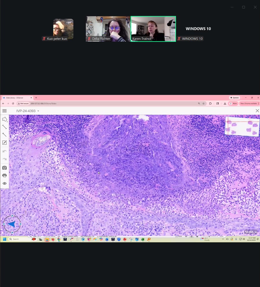
We’ve also got this really thick, richly cellular-like noid to interface pattern. And it’s very histoacidic with that fine dusting of melanin, and very fine faint granules within the cytoplasm. And that is very characteristic for VKH.
🖼️ [00:00:26]

Sometimes it can be a little pyogranulamilatous like we’re seeing here. This has a surprising number of neutrophils in it.
🖼️ [00:00:33]
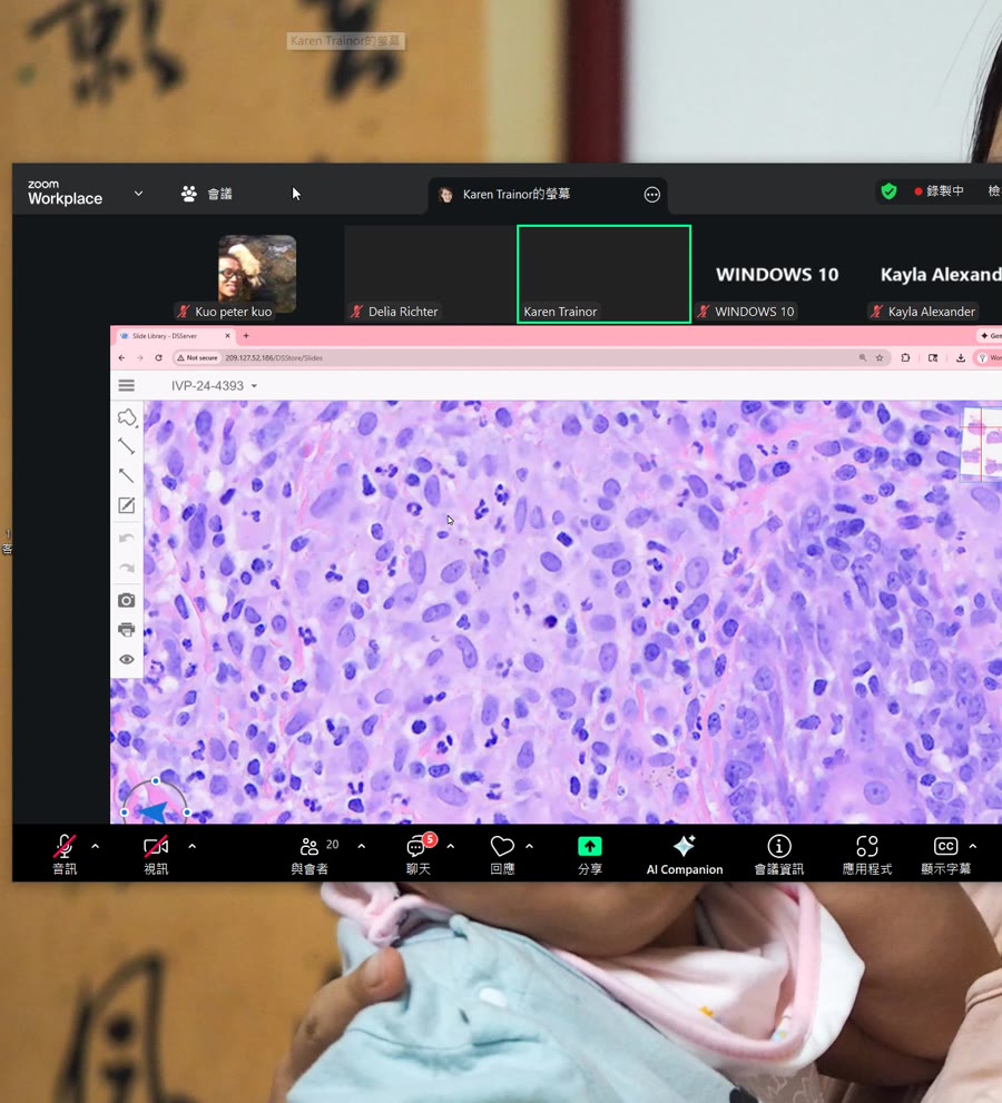
And then other times I have seen VKH present, it’s tracking along at Naxil structures here. I have seen it present almost folliculocentric without the lichenoid or interface band, but it was concentrated around the hair follicles. So that’s something else to keep in mind if you’re reading biocular. or if you have, or if you’re a clinician seeing patients and you see the lukatricia or
🖼️ [00:01:00]
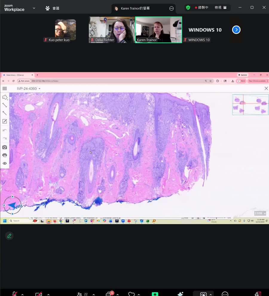
leukoderma, might be something to consider, especially if the pathologist is considering, like, maybe neural diseases or periodnexel causes of inflammation and they don’t list BKH. If it’s the right breed or if you’re concerned at all about ocular lesions, VKH can rarely present that way. Yes, Discoid lupus would be another excellent differential here. It’s the right distribution, especially being mostly facially predominant. But you’re right, it wouldn’t be this dramatic. It’s usually more toned down than this. Any questions on this case? All right, I will go back to our presentation here. I can do this. And you guys had excellent differentials. We identified this is hair and skin from the face. We have this lichenoid interface band. It’s very thick, also extending around it nexus structures. Sebaceous glands are retained. And we have the patchy loss of the granular cell layer, the paracarotosis with extension into follicular osteo, and also the serocular clustered with the coxye. So we have VKH, probably a component of zinc responsive dermatosis, and secondary bacterial bioderma. three diagnoses here. So some cases can be very complex and very layered, and it’s important to keep all that in mind and not just stop once you see one diagnosis or one that jumps out at you. Sometimes they can be nuanced or more complex. So we have looked at all of this. This is the kind of pyrogranulomitus area of that lachenoid band and then just showing that fine. dusting of melanin in the macrophages and those histocytes. And just to point your attention again to the leukotrichia, that is a really key feature to look for to help make this diagnosis. Because once you get that in your description, that helps you separate out from DLE where you might get leukoterma, but you’re not necessarily going to get leukotrachean. So that would really be very helpful. All right. Moving on to our next case. This is from a cat. This is a really cool case from earlier this week that I wanted to share. In all my years of reading biopsies, I haven’t seen this in the U.S., so kind of fun. So let’s see. All right, we have a six-week, two-and-a-half-year-old female spayed rag doll. Beautiful cat. My name. next cat will be a rag dog because our current cat is my husband’s cat and I’m very jealous and I want my own cat. That just prefers me and I want it to be a rag dog. I’ve always had free cats, but all right. So this poor kitty has had a six-week history of progressive, severely, very severely prudic, crusting dermatitis affecting the nasal bridge, the pin of margins and the chin. She’s also had a consistent submandibular lymphaticis. and she has systemic signs of fever, soft stool, and some diarrhea. There are other cats in the household, but no other cats are affected. So the diagnostics that were done so far, you can see all the cresting here on the margins of the pina, on the back of the pina, negative Woodslamp exam, negative dramatified PCR, negative fecal antigen flotation, negative FELV FI, tests. CBC, this clinician did a phenomenal job, absolutely spectacular workup on this case. This was from a general practitioner. A CBC that revealed a mild non-regenerative anemia with a leukocytosis, a left-shift neutrophilia and eocinophilia. Chem panel showed mild hyperglobularemia and urinalysis was unremarkable. No ectoparasites have been seen at any visit. So I asked, you, what are your differential diagnoses? These are the pictures of the time of biopsies. Let’s just imagine there are gloves in this picture. Yep, still suspicious of parasites. Which parasites would you be suspicious? Sarcoptes, that’s a good one. What other differentials would you have? Notoed eidres, but other cats unaffected and young cat. Yep, pampagus, foliaceous, excellent. Pemphagus. Mm-hmm. Always a popular choice with cats. All right. So, Leishmania. Excellent. Yes. Oh, yep, because I mentioned geography. Yep. Pox. Ooh, that’s a good differential, but no upper respiratory information. Correct. Yes. Great differential. That was not on my radar, but I’m glad you mentioned them. In the EU, it would be POTS. Oh, I’m learning something new. Thank you for them. all right let’s go to the histopath because that’s the most fun. All right, let’s see if I can successfully switch over again. All right, so we don’t want this one anymore. We want, I can get my case numbers here. All right, let’s do this slide. So where is this biopsy from? What part of the body? Do we see subcutus? Face, very good. No? Well, yes, we did see a lot of punches from the face and nose. I did show you that in the last time. That is fair. Yes. So haired skin from the face, excellent. Yes, very good. And the sebaceous glands are quite large. On the face in cats, the sebaceous glands will be pretty big. What draws your attention already at this low magnification? mites mites yes good times all right let’s take a closer look shall we gotta love them so sometimes histories can be a little misleading so you have to still think broadly with your list of differentials even though and I trust this clinician they’re an excellent clinician they’re very thorough very methodical in their workup do like everything they go by the book. Sometimes there can still be curveballs because this clinician also sent me clinical photos so I could shop the photos and tell him where to biopsy, what biopsy technique to use, where I thought we’d get the most bang for our buck for diagnostic samples. And we were both leaning towards pemfagus, most likely, with all of that history. But plot twist. So, yes, we’ve got mites. So we’ve mentioned, there’s been mention of a couple different mites. Which type of mites would you consider here? There is sarcoptes, no-o-edris. You might also consider potentially demodex, no-o-edries, okay. Soroptes, otodectes, okay. what are some clues here to help us? We can measure this mite for a size approximation, but what’s a clue that will help us between all these wonderful different mites that you guys are listing? Burying versus not growing, right? Excellent, not in false. These mites are up on the surface. These are superficial. But more than that, where are they on the surface? They are sort of in these tunnels of paraccharacteri paracarotosis. They’re in the stratum corneum. So this is all nucleated keratinocytes sort of enveloping around the mite. So the mites form tunnels. Exactly. Yes, these are burrowing and not Demodex due the shape. Demodex would be much flatter and don’t really have these ridges on the cuticle. And this is the chitinous exoskeleton all around. And then we have the muscle here. These are some internal organs. Don’t ask me to tell you which ones. So yes, we’ve got mites. We’ve got all this acanthosis here on the surface and paracaritosis. We’ve got some peribascular to interstitial dermatitis. Human stuff. In this case, I asked the clinician. This is one where I stopped. I didn’t even finish writing the report or photographing it. I picked up the phone and called, was put on hold, so then sent him an email in response to his email and changed the headline to urgent because he’d already started treating for penfegas because the cat was so miserable. He had written and said, can I start treating for penficus? And I usually say, I’m not the most appropriate person to ask about treatment. I defer to the clinicians for that. but until we could get him a clinical consult, he said the cat was just epically miserable. I was really suffering. So he did start steroids. But then he also contacted the owner because we were both surprised. My first thought was maybe sarcoctes or no to Idris because they both look very similar. They both are in the same family of sarcoptidae. and they both tunnel and form a lot of paracarotosis and acanthosis. And you tell them apart, in part based on the signalment. So cats, it would be more likely to see no to Idris, although sarcoptees have been reported. I don’t know if that would still stand up. That was a pretty old article, as I recall, but there were several cats that had sarcoptes. If anyone is more up to date on their mite literature than I am, please feel free to chime in. But you go based on the size of the mite. And so the clinician, while I was measuring trying to get dimensions of these mites to confirm if it was sarcoctes or no to Idris. The clinician was on the phone with the owner asking if they had any lesions, like just confirming because he didn’t remember them mentioning anything like that. but they said no lesions on the people. So he is very surprised by this, as was I. But we can take some measurements here that we measure from here to here. It’s only 101 microns. So that is fairly small. That is less than about 250, which I would expect a larger mite for sarcopty. So the smaller one, I would expect no to eG. So this was just a fun case I wanted to share with you because it’s not something that I get to see very often here in the U.S., I guess fortunately. But there are pockets of no-do-ydris in cats in Florida, in the Pacific Northwest, and I believe in Texas as well. Maybe some other locations sporadically, but this was a case of Notre-Ajuris. Just look at this other sample as well. Also from the face, this might be the junction there with the nasal planum because we have sort of this transition from haired to non-haired skin. And what are these structures right here that we’re looking at? Does anyone know? The pathologists will know. Nerves, yes, peripheral nerve fibers. It’s a very highly innervated area on the base. This is an axon that’s coming right out at us. and cross-section. Very good. All right. So then we have this perivascular to interstitial dermatitis, mostly mass cells. Maybe a few eosinophils in there. They’re kind of hard to identify. Just a couple. So not as eosinophilic as I would have expected. If we didn’t have mites, if we were unlucky, this might just come across as being an allergic dermatitis. But if we’re lucky, we happen to capture the mites right here, I’ll measure this one. This one looks bigger. This one is 208 microns in greatest dimension. So still smaller than 250. Gorgeous. Beautiful biopsis. Well done to the clinician. So that would be a tip for the general practitioners. Or anyone that’s looking for optimal results on their skin biopsies would be to take clinical photographs. and share them with the pathologist if you’re at all uncertain of what technique they might recommend or where they want to biopsy so that you guys can work together to optimize and make sure you get the best samples. Because there’s nothing more frustrating than someone taking biopsies but not capturing the most optimal area to get a definitive diagnosis. There are some needs and feels in here. any questions on this one? We’ll go back here. So this is another sample from the face. We have all our acanthosis and all our fragments of mites on the surface. And also within these tunnels of paracarotosis, this tunnel is broken open. Yes. So beautiful example of that. We can see them in cross-section. We saw this section already. These are the mites with the paracarotosis and inside of these tunnels. So they’re burrowing within the tunnels and they’re not in the follicles as you’d expect with demodontes. All right. And this one I measured as 220 microns on my regular microscope, not on the scanning, digitally scan slide. This is on the glass. Alright, so this is very rare in cats, at least in my experience. Please let me know if your experience is otherwise. otherwise. And there are some pockets that are found in Florida, and this cat, sure enough, is from Florida. The Pacific Northwest and Texas. Possible sources of exposure can include squirrels, rabbits, raccoons, and bobcats. Noto-Egris mites are contagious and spread via contact. And if one cat in the household is affected, It should be considered that all cats in the household should be considered as infested. And it is a member of the sarcopta day family of mites. So it has a very similar life cycle to sarcopty scavii in dogs and wildlife and people. And we’re familiar with that perhaps a little more. And so there’s a lot of paracarotosis with numerous tunnels that contain the mites. and they’re up to about 250 by 150 microns. Bigger than that would fit more with sarcopics. Smaller than that would fit more with nootidris. And nootidase also is associated with lymphadenopathy. And the distribution of the lesions in this particular case, being over the nose and over the pina on the face and being extremely preretic is another key diagnostic feature. All right, any questions on this case? And then I’ll take a quick poll. I don’t know if you guys want me to keep going. I have two more cases or we can stop here or what would you guys prefer? All right, more cases. Interesting. All right. Excellent. All right. We’ll carry on. If you have to go, that’s fine. But I do have a couple more prepared. So we’ll take a look at those. Thank you for the feedback. I appreciate it. All right. So our third case is from a dog. That was our only cap today. But I hope you’ll find this case interesting as well. This is from a three-year-old female spade golden retriever mix. She is quite adorable, but she’s very dainty and petite for a golden retriever mix. I don’t think I’ve ever seen one this small. But sure enough, this is what she was listed as. and she presented for a recheck exam for some new postules and crusts that had developed over the head, neck, thorax, flanks, abdomen, and paws. And this clinician did cytology and found neutrophils and coxye on cytology and a few acetylidic keratinocytes as well. So the dog was started on cephcdoxomy and to address the pyodermy. and then we’ll see what happens next. You can start to see some of the postules here on the skin on the slides. All right, so a week later, the lesions have progressed and are impacting more of her body, and she overall seems very lethargic and very pritic. On physical exam, there are numerous intact postules. You can see them right here. These are beautiful. I should zoom in even more and crop this image, so you can see it there. but I don’t know if I don’t know if I’m not letting me zoom in. So there were numerous questions. And then they had multiple adherent yellow to gold colored crusts. We’re always on the lookout for honey colored crusts over the neck, dorsal thorax, flanks, and over the dorsal surface of all paws. So my question to you, what are your differential diagnosis? I’d also point out, yeah, pempel. I guess foliaceous, excellent. Yes, yes. I would also point out her coat isn’t looking so hot. Golden ichthyosis, yes, non-epidermalytic icktheosis as opposed to epidermalytic. Good thought. Lymphoma, good differential. Lymphoma can do anything at once. This is young, on the young side for lymphoma. I’ve seen epitheliotropic lymphoma in dogs as young as four, and it was actually. from this practice a different doctor. And that dog, the main clinical finding was leukotrichia in that four-year-old dog. Yes. So typically for lymphoma, I start thinking of that when dogs are nine years of age or older, but I still keep it on my radar, even for dogs younger than that, or animals of other breeds or species. if they’re younger, just because it’s kind of a wild card.
🖼️ [00:22:47]
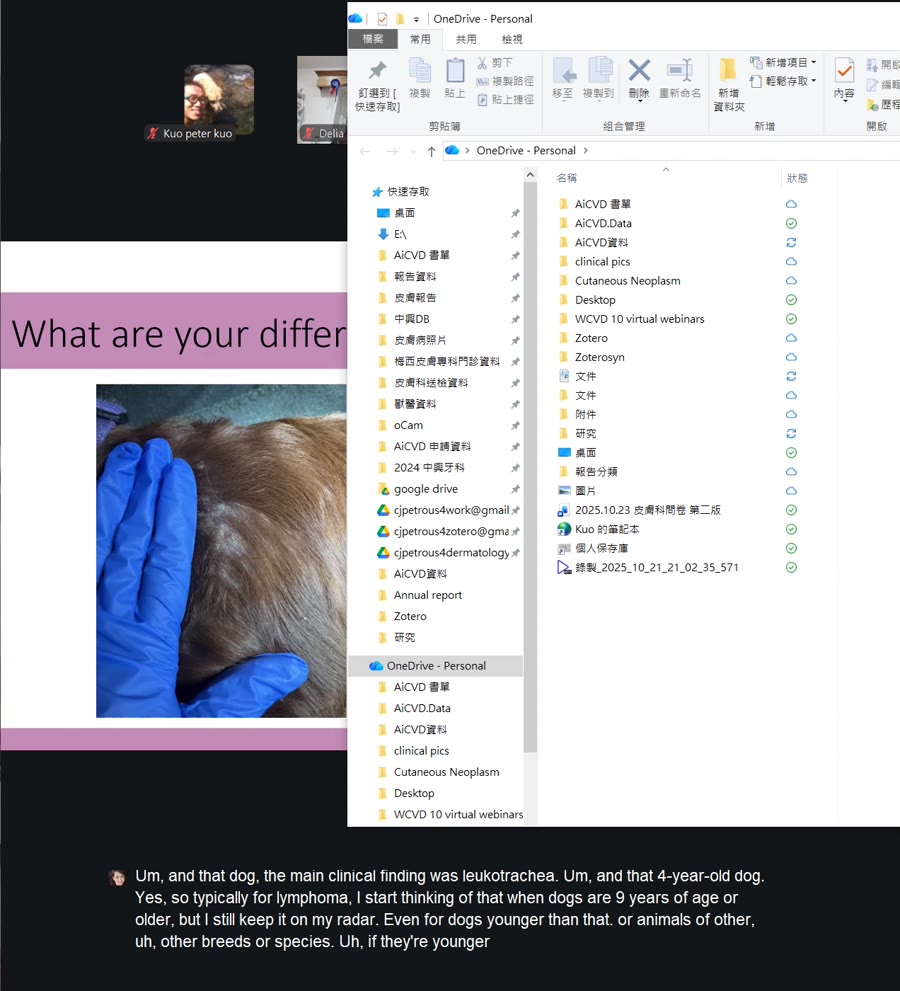
So certainly something to keep in mind. But her coat isn’t looking so hot. So there is something else going on. So we’ll take a look at the slides and see if we can figure out all the layers of what’s going on here. So we want, I’ve got to get my numbers out. We want 577.30. Oh, now these biopsies look different. You can already appreciate that these are not from the face because at low magnification, we have plenty of subcutus, and we have our underlying muscle down here. The muscle is not intermingled with the genus. So, oops, all right. If we zoom in, what have we got going on? What pattern would you describe the sense? This dog read her textbook. So pampagus was mentioned as a differential. This indeed is a broad subcourneal postule. And we do have neutrophils in here, maybe some eosinophils as well, and some acanthalytic keratinocytes. So this would certainly fit with pempfagus foliaceous, and I’m not seeing colonies of coxye like we saw in the first dog. So I think that antibiotic she’s been put on is helping to knock down the pyoderma. So there could still be a little bit left, but this is quite a lot of acampalytic keratinocytes, more than I would expect for pyoderma alone. And then there’s a peribascular interstitial dermatitis. But she was seen by a dermatology resident, and she was seeing for a recheck. So she must have something else going on. It says, haired skin. Is there anything that you would expect to see that you’re not able to identify? I’ll go to some other section. No sebaceous glands, yes. Well done. Okay, I have to bust out the gold stars here. Well done. Yes, very good. So she is being treated with cyclosporin for her sebaceous adenitis, and she still developed these lesions of pentagus. phylaecious while on the cyclosophobic. So she was in need of another biopsy. She’d already had one previously with another, with her mentor at the practice, who was a dermatologist. And he had submitted biopsies from this dog previously to diagnose the sebaceous adenitis. But now she’s got a new
🖼️ [00:26:04]
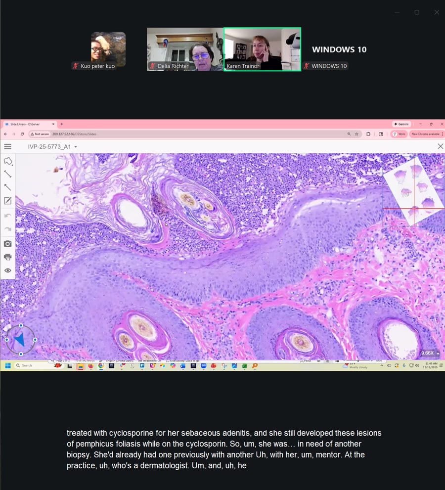
problem and it’s this pampagus violation. So again, just on the theme of dogs can have, or cats can have multiple disease processes at the same time. And it’s really important to work with someone that is very like dedicated to germ path and doesn’t just stop immediately with one diagnosis because sometimes there can be two or three. Don’t, off the top of my head, it’s very hard to think of a case that had more than three processes at the same time. But just something to keep in mind. And for board exams, for pathology, for, I’ve heard for dermatology, I don’t know if it’s still the case, but that they will sometimes put two diagnoses in a slide or image, say squamousal carcinoma and demodex in the same image. So two for the price of one. I know at least anatomies. pathology boards were notorious for that. Very good. So something to always check for on haired skin biopsies if you’re reading germ biopsies is always checked no matter what is going on. Always look for sebaceous glands. So they are absent here. Where on the body would you expect to see very sparse sebaceous glands, which might be harder to assess. So the sebaceous glands are really prominent on the face, especially in cats, especially on the chin. But where are they sparse on the body in dogs? Pena, they’re pretty small there, but also in the ventrum. If you have a dog and you’re in practice, especially in general practice, and you suspect sebaceous adenitis, and you are going to take biopsies, the best place to biopsy in dermatologists and germ residents, please chime in and correct me if I’m wrong. Typically, we recognize. recommend biopsying along the dorsum, especially over the dorsal shoulders, because that’s where sebaceous glands are the largest and most highly concentrated. So if you’re trying to make a diagnosis of sebaceous adenitis and a dog, those are the best areas. And there is a form that OFA uses, the orthopedic foundation of America. They are sort of the clearinghouse for poodles that are being screened for sebacea adenitis. And they have a nice handout that they give clinicians that tell them where to sample, and they recommend over the withers is the best area to sample. This is a nice, beautifully oriented specimen. We have the broad subchoreal postule that spans across hair follicles. Another differential I might have for this would be superficial spreading pyragerma because that stratoncoran is lifted so nicely. but I’m not seeing much in the way of bacteria here. The neutrophils are very well preserved. I’m not seeing nuclear streaming. And we’ve got nice acanthalytic keratinocytes and even some eosinophils. So once I start seeing eosinophils in the postule, I start thinking pentfagus, at least in dogs. And then we also have the acanthalysis here. You can see the cells on zipping and becoming detached. from each other. So there’s that and then in profile at the level of the isthmus there are no sebaceous. They are down and out. All right any questions on this case in humans they see acampillic keratinocytes in dermatophytosis why less of that in dogs? That’s an excellent question. I don’t know. I certainly see a couple of of cases like maybe one or two a year of postular dermatophytosis and dogs, but that tends to have more follicular involvement. And then the acantholysis is not in the handful of cases I’ve seen. It’s not something I see often. The acanthalysis is not as decadent as in this case. Like here there’s a lot of them. You can really, you don’t have to search too hard to find them. In the Pusular Dramatophycotosis cases I’ve seen, they’ve been more sparse. But I don’t know if that’s just the luck of the draw. I think during a Davis-Thompson Foundation lecture, Verena had mentioned that she does not go based on the number of acacanthalytic keratinocytes. So I’m going to pay more attention to that to see if that’s, if that, you know, holds up because that may. may well be true. But yes, and I have seen one or two, well, one that I know we have confirmed cases of dermatophytoe, postular dermatophytosis in a cat, which I’ve read about but never seen for myself. So maybe another time we’ll do that. All right. Let’s see. Let’s go back to our slides. So you guys did a great job.
🖼️ [00:31:52]

So this is Penfegis booleatious with our broad subcourneal and it even extends down into the follicular epithelium and the perivascular interstitial dermatitis with some edema in the superficial germis and then the lack of sebaceous glands. All right and then in the postural there’s plenty of acanthalic keratinocytes and the neutrophils are well preserved. Again, you’re going to look for the honey-colored crusts in dogs. That’s really like the quintessential clinical finding. You can see a mix of well-preserved neutrophils and sometimes eosinophils as well. And this clinician, the resident, she did an excellent job. She did her cytology. She did see nutrifil. She did see acynthetic carotinocytes, but she also saw coxcine. So what are the top three causes of acanthalytic keratinocytes. We’ve kind of already talked about them, but just for anyone that’s in general practice or any residents, it’d be good to review. So bacterial pyoderma, hemophagus foliaceous, and dermatophytosis, or your top three causes of acanthalicis. There’s another one that’s more obscure. If you’re a young Bernie’s mountain dog in the upper Midwest of the United States. There is now an emerging disease that I’m working with a lot of colleagues. We’re all collaborating from all different backgrounds on a project to study Darius disease in Bernie’s Mountain Dogs, young Bernie’s Mountain Dogs. We’ve got genetic testing that identifies it, but they get a lot of acetylysis, but they have it without the neutrophils. So there will be no neutrophils. introphiles with it. It’s just pure acanthalysis. And they refer to the acanthalic carotinocytes as core roms. And it’s a fascinating disease and maybe I’ll show one of those cases another time. But anyway, those are, that’s a more obscure, more trivial cause of acanthalysis. I wouldn’t say that’s a common one. But just reading routine diagnostic cases, those would be the big three I keep in mind. all right so i think we have times go through one more case quickly if you guys are up for it yes no all right okay this is a fun one that i only get to see like maybe once or twice a year um okay and then the sebacea adenitis the key findings you guys are probably all all pros at that um since this dog had two different diseases um all right so case number four this was another case from I think just earlier this week. Yeah, I guess it’s Friday. It was earlier this. It’s been a long week. All right. So, yes, Bejohn and Havonis too. Thank you. I need to update that. Thank you. You’re correct. Absolutely right. I will make a note of that. Get that to do that. Noted. Thank you. All right. So our last case is from a dog. And this poor dog. Snoop is a six-year-old male doxen, and Snoop Dogg presented for a recheck exam. And a few months ago, he had three seizures in one day and was started on phenobarbital. Then he started to gain some weight after starting the phenobarbital, and they checked a total T4, which was found to be low, and so then he was started on Levo-Thiroxiax. Then within the last few days, this had been going on for some month. He broke out with lesions on his nose behind his ears involving the axilla and abdomen. And he was also started on an antibiotic and there’s been no improvement. So this is coarse new and we see some erythematous, nodular, macular lesions on the nose, on the bridge of the nose and the muscle here. And then he’s also got lesions on the penna. So on physical exam, the clinician noted multifocal firm erythematous nodules on the coddle basis of the peni, multiple firm erythymidus nodules under the axillary, and multifocal erythematous firm nodules on the ventral abdomen. There’s also like kinification with erythema and crusting on the dorsal muscle and mild interdivocal. digital aerothene. I think there’s more clinical photos. I would point out the excellent use of gloves by this clinician. Also, great photographs, you know, good lighting. It shows that there’s, you can appreciate there’s some thickness to the lesion, good resolution, but gloves, importantly. So I do like to mention that to clinicians, because if it was that poor kitten that we saw from Florida, I would want you to be safe. I wouldn’t want anything to spread to you. All right. So you can see the lesions on the abdomen as well. They are certainly erythematous. So what are your differential diagnoses? Drug reaction. Eosinephilic foroculocyluses, certainly. Excellent differentials. Erathema multiforme. Yes, that is probably my top differential for a dog on seizure medications would be aerothema multiforma drug reaction. It’s probably the top plate that I see, or dermatophytosis. Certainly, it’s a possibility. Superficial necrolitic dermatitis or hepatocutaneous syndrome, that’s a good differential. Certainly being on phenobarbital. Excellent thought. So let’s go to the histopath because that is interesting. This is not something I get to see very often. So, this is not our case. We’re just going to focus on the histology. Which one do we want? I think they both show the same thing. We’ll take a look. All right. So we have haired skin. Okay, I’m going to make everyone dizzy. I should have warned everyone to take their drammamine with the Zoom link. All right. So we have hair and skin, not from the face because we have subcutus here. But how would you describe the district? This is just artifact for anyone that’s not a pathologist. It doesn’t look at slides all day, so just regard that. It’s just a little floating piece of tissue from the water baths. Let’s look at this one. So I would describe this distribution as perivascular to interstitial and bordering on diffuse. And we can see at low magnification, we get a glimmer of what the collagen should look more like. It should be more compact and eosinophilis. and eosinophilic and dense. And most of it is pretty interspersed or spread out and sparse. There’s a lot of clear space. And then also some almost, you can get a hint of like these pale blue wisps. So there’s edema and then over here you can see it more clearly. What is this material? Do you think? All right, Musin. Excellent, very good. We have Musen as well. So what are some, yeah, mix edema. What are your differentials once you start seeing a lot of mucin in the gen? Not a lot. Another short list. Hypothyroidism. What about a breed specific? Sharpay mucinosis, wonderful. There is one more even obscure breed, even more obscure than Sharpay, that can also develop mucinosis as an incidental finding, not related to hyposyroid. It has a black tongue. Chow Chows. Yes, very good. I think everyone said at the same time. Okay, wonderful. It’s round sound. Yes, so Chows can also, I only learned that recently, like a year ago. And then what other diseases can cause lot of eosinosis? What if it were in a cat? What if this were a skin biopsy from a cat? And we saw a lot of mucin. Cats tend to get hypersticinitis. thyroidism instead of hypothyroidism. So in cats, if they have eosinophilic, they have a whole range of eosinophilic skin diseases, but if they have like an eosinophilic plaque, you can see a lot of mucin deposition with that within the apodermis or even in the dermis. You can see quite a bit of mucinosis. Musinotic neural folliculitis of cats, excellent differential. Sometimes with eosinophilic granuloma, that’s absolutely right. So we’re seeing here, I’m zooming in, we’ve got these eosinophilic granules in the cytoplasm. We’ve got a lot of eosinophil. This is peribascular to diffuse. Eosinophilic, maybe some mass cells, some mass cells mixed in, but a lot of eosinophil. I would also say mass cell tumors are a possibility for developing musin. Yes. So like urticaria pigmontosa or cutaneous mastocytosis, you might see mucinosis with that. And then no one’s mentioned this condition yet in docks. We’re seeing like a diffuse pattern of eosinophils with edema and mucin. Let me show you some other areas that that’ll be a clue to give it away. Let’s look right in here in the mid-dams. What are we looking at here? It’s a bunch of Asinophils, they’re swirling around collagen bundles in the dermis, and they’re starting to degranate. So we’re starting to get a collagen flame figure happening here. So yes. So that cutaneous, yeah, an allergic response. So this is eosinophilic dermatitis, canine eosinophilic dermatitis or Wells Lake syndrome. I don’t know that I would call this C-A-E-E-D-E. In particular, in this case, this is not associated with GI disease in this particular dog. But this is, I think, on that spectrum. I think there’s still a lot that we need to learn, or at least I need to learn, about eosinophilic diseases in dogs. Yes. And maybe the terminology is updated from what I studied when I was a resident. This canine is synophilic dermatitis with edema. I think they call it like acute arithiderma or something like that. There’s the A in there. And you can have some vascularopathy as well. All these cells are migrating through the vessel ball. So this is a hypersensitivity response. There is thought to be a seasonal component to it, which I think is something that might differentiate it from that C-A-E-E entity, but please let me know if I’m mistaken on that because I’m not necessarily up to speed on every manuscript. So this is the dermal mucinosis and edema with the diffuse inflammation, and you can see these collagen plane figures starting to form. And we also have eosinephalic follicitis happening as well, which I don’t think is commonly the same C-A-E-D. So this is the eosinophilic folliculitis. And that fit. Someone mentioned that before. Eosinephylic follicillitis and perunculosis, that region on the muzzle, the dorsal aspect. That fits pretty well. Collagen-in-flin-figer getting started. Not quite an eosinophilic granuloma, but hard to say what some of these cells are. They’re probably degranulated eosinophiles, but could be massans. This is also called Wells Life Syndrome. It’s a relatively rare diagnosis, and this dog read the textbook. It does tend to occur in late summer or early autumn. This dog is also from the southeast of the United States. And it was just recently, so it was kind of, well, maybe early autumn in the south, in the deep cell. So possibly due to arthropodice salt, which would fit with the aesineophiliculitis and foronculosis, which is also thought to be arthropodice salt, or, oh, looks like someone’s helping us out here. Thank you. I appreciate it. So check in the chat. There’s a link to an article. Thank you, Nicoletta. Well done. Appreciate that. So seasonal alertic reaction or a drug reaction. So this poor pup has a lot of potential triggers. Could have been arthropartisols, especially that lesion on a muzzle. Could have been an allergic process. Or it could have been a drug. reaction because now she’s on all these different medications. The lesions do not blanch with dioscopy. And we went over this case in rounds earlier this week with the groups that I work with, and the resident did confirm that the lesion did not blanch with dioscopy. And they tend to be generalized, but it may affect the ventral abdomen, thorax, base, and peni, which is exactly the distribution we had in this case. Pyritis is moderate, typically, and, and the end uproids. and lymphadenopathy is common. In humans, there’s typically a peripheral eosinophilia, but that’s not found very often in dogs. Hystopath is required for diagnosis, but the clinical lesions can be suspicious for it. On histology, you will see some acanthosis and then moderate to severe, superficial to deep because this was pretty diffuse, peribascular to diffuse, eosinophilic. dermatitis, and there’s edema and mucin. So I would think it’s mostly related to that. I need to get more follow-up from the resident about the thyroid. But usually with the hypothyroidism, it’s very rare for me to see like this degree of mucinosis. It might be like just a small wisp of it if it’s due to hypothyroidism. But usually those dogs will not have concurrent inflammation. It’ll be a non-inflammatory, non-scaring alopecia, and they’ll have follicular atrophy. And I did not appreciate follicular atrophy in this case. So I’m leaning towards this eosinaphylach dermatitis, which is rare diagnosis. So any questions? Thank you so much for sharing that manuscript. I really appreciate that. All right. Well, I think that’s all I’ve got. If you have any questions or think of anything, go ahead and send me an email. I think we’ll try and have a reporting of this available if you’re not able to attend my. Thank you so much for your participation. I hope these were interesting and hopeful. It was a great discussion. I really appreciate everyone’s attention. And please let me know if there’s any other topics or cases you want to see in the future. I’d be happy to organize them. all right well thank you so much everyone have a great day i’ll see you next time
🖼️ [00:50:12]