2026.01.23 ECVD HistoRound(自動整理)
由
錄製_2026_01_23_00_01_53_115.mp4自動產生:faster-whisper 逐字稿 + ffmpeg 切片截圖。 ⚠️ 這是機器自動整理的原始版(英文逐字稿可能有小錯、切片圖是自動抽的)。看過後再自己濃縮成重點、補中文。
- 已人工校對/濃縮
🧩 內容(切片 + 逐字稿,依時間交錯)
we could not understand which was the pattern because nothing seemed very special to me. Something that at low magnification I saw, but I’m not sure, I wanted to ask you this, is because, for example, in this section, yes, this one, like there is a part of the dermis, which there are no follicles at all, and then all of them are, why is this happening? I think it is a cat artifact. Okay, perfect. I have no other reason. I would not know because this is not, I mean, one reason why you should have this could have this appearance of hair follicles here would be scarring, right? However, this is not a scar tissue. No. There is no increase of fibroblasts. So I would say that this is a normal thing, normal dermis, let’s say. So also if you look at the lower part, the hair follicles are just fine. Yes. So I think it’s just a catarctic cuttacus strange catharticotis. Okay, perfect. Then, well, I started at the top which I saw this moderate to severe diffuse basket with ortho-caratitis. This is a very nice example. example of a very nice but very prominent orthokinatonic basket swim because normally you don’t see it is perfectly okay and then I wrote moderate epidermal hyperplasia and then I was like what is going on I’m not seeing very much then I was like where is the inflammation I’m not seeing a lot of inflammation going on So I was like, but then I saw a lot of blood vessels. And I believe a part of this infiltrate is around these blood vessels. So I wrote pattern perivascular. Yes. Okay. Perfect. Then I start to see a lot of eosinophils a lot. I see some eosinophils. Okay. But when I see eosinophiles, I’m like, is this really a eosinophil? Are we sure or not? I have to say I didn’t pay attention too much to the Yusufeld, so I think we should try to find them. Do you remember where they were? Some of them were mainly inside of the… Yeah, something like this, for example. Yes, inside of the blood vessels. I see a lot of them. Yeah, yeah. You know, I’m not. even sure if these are eosinophils or neutrophil the nucleus is very convoluted so i would say that this is more more more likely a neutral feel rather than yeah okay okay yes i was uh yeah and until you see them in the vessels i would not i mean pay attention yeah no the majority of And these, to me, they are like here or here they are inside the vessels. The blood vessels. Yeah. Okay. Okay. And then in some of the samples, not all of them, but there are some mild to moderate edema. Yes. Yes. Yeah. Yeah. And this is mainly what I saw because more or less it was normal, so I was normal. like not sure what I am supposed to see here. Okay. So yeah, in these ones, I think you have more of edema. Yeah. Yeah. Yeah. Yeah. Yeah, for example, root. Yes, more edema. That was like, and also on some blood vessels, they are like a little bit dilated. I’m not sure if this. Yes, yes, yes. Sure. There is atyremia, there is congestion. Congestion, yeah. It is called sometimes leukostasis. So you have a lot of leukocytes inside plugging, kind of the vessel. And I agree with you, totally agree with you, that here there is a lot of edema. See how separated are the collagen bundles? Yes. Yes. I was thinking something like, but I’m not sure, like something eurtigarya, something like this. Yeah, it could be. No, no, I agree. I agree. There is something else, let’s say. The hyperkeratosis is sometimes even paraccharacritic in the hair follicle. Ah, these are you don’t show. And you also have some. So if you look at this, no magnification. I totally agree there is nothing very much, but you have the very kind of, um, a little bit strange epidermal hyperplasia with hyperkeratosis. You see that there is some kind of, um, poppylated appearance of the epidermis. So yes, it is a dematose. I agree with you, but it’s kind of chronic. Okay. Right. Okay. Okay. Mm-hmm. Sorry, no, no, go ahead. So it’s chronic because I see like this hyperplasia, no, which is a chronic, yes? Yeah, there is hyperplasia. And this hyperplasia is not the most prominent case, I would say. and also this is something that was given up as these so we are trying to find that there is this kind of festonin of the epidermis you see it’s not just the epidermis that is irregularly hyperplastic there is some repetition in the hyperplasia you see now that you mention it yes i see so there are these they are like buds of epithelium that are like regularly doing this hyperplastic appearance and I have to say and I have to be honest this was given as malacetia what I did and if where is it yeah exactly if you go and look for it there are a couple of my God, didn’t see it. No, but I mean, I have to say I mean, I look at them after knowing what it was. And indeed there are a couple of these. There are also I can’t find it now but there were I think In one section, they were even in the hair polychord. And so maybe this is one, I’m not pretty sure. It’s a second. Claudia von Chanier used to say that if you, because you know that if you find a couple of malacetia on the surface, it’s okay, can be… It’s normal, yeah. Normal. You see, I mean, here and there there are some yeast. But I had the impression and I can’t find it again. These are probably just bacteria. That were also in the lumines of the follicles. If you find in the lumines of the follicles, of course, it’s more, let’s say, heavy in terms of interpretation. Okay, here, for example, you have them also in the lumen of their follicles, right? Okay, yes. I agree with you, there are not so many, but there are some of them, okay? Okay. Otherwise, I agree with you, the pattern is one of a perivascular superficial dermatitis. In this case, it’s not even very severe. Yeah. The diagnostic keys isologically are these budding of the epidermis and often, and sometimes it was prominent, you have severe spongosis or, I mean, you have some spongosis and lymphocytic exocytosis. That often is patching. Okay. Okay. Okay, perfect. Thanks. Kiarra, can I ask you a question? Yes, of course. Because I thought I saw perinuclear vacuoles in the stratum granulosum, and that really brought me to another diagnosis. Perinuclear vacualled, you thought that they were inclusion bodies? No, I thought it was ictiosis. Aha, okay, okay. Yes, let’s see. And I saw them somewhere on the right side of one of the slides. And I don’t remember where anymore. The right side is here. It was the, how do I say that? It was the middle and then above. This one? So, oh, I have to check. Where is it? Oh gosh, I turned them, of course. For the, I mean, these perinuclavacos that you are describing are described, sorry, in the non-epidermolytic heiosis of the golden retriever. And I have to say, I have a hard time to think that these are in way characteristics, or histologic key of the osteosis, the non-epidermalytic ethyosis, because, and this is an example, I have the impression that you can see these perinuclear vacuoles, even in other diseases, and this is an example, if we find what you saw, but sometimes you see vacuolated superficially, spines, keratinocytes, or even in, normally you see them in the superficial layers. What to me, if this was to be, let’s say, counted or interspreaded, what it would speak against ichtiosis is that the keratin is very, basket weave or lamella, whereas in the iosis of the golden retriever, usually it’s a little bit more compact and it’s very eosinophilic. And usually the dermis is really clean, totally clean. Here, I agree that it’s not very prominent, but there is a a perivocular inflammation and there is edema. Mm-hmm. Okay. Does it make sense? Does it make sense? Yes. Okay. Wait a second because I… Let’s see. Okay. Just one second. If we can… You see my screen, right? So you do, you see whatever I do. Yes. Yes. Yes. I’ll try not to go on my… Okay, so this is a case of ecteosis of the golden retriever. Okay? So you see how different the keratin is. It’s very eucinophilic and very tight, very compacted. Even if it stays lamella in some ways, it is very compacted. Let’s put it in comparison. Especially if we go like in the places where it was ortho-keratotic, of course the difference is very marked, right? Can you answer to me? Yes. Yes, but all is more pronounced in the right one. The epidermis is also more blueish, but violets than on the left side. Yeah. So it can be also the staining that is more toned on. Most like more likely, yes, for sure there can be one of these components, but like here, for example, you have no problem imagining the scales that you see microscopically, right? clinically. Okay. And one typical thing is really the the staining, the color, it’s really pink. Okay. And then there are these lesions, these vacillation of the superficial keratinocytes. That again, it is described like here, for example, it is described in the books, in the, in the papers, even in the paper from Elizabeth Walden. I am not sure if it’s just really typical and or let’s say exclusive of the of the of the eidosis. I don’t think we could see the who was speaking, sorry. I don’t think it was. It was. It was. It was. or Nicole that was talking about this perinuclear vacillation. Yeah. Was Nicole, right? Yeah, it was Nicole. Yeah. Did you, by the in the meantime, did you, did you check where you saw this cell? Yeah, I think it is, when you look at all the different slides, then I think it’s the underline and then it’s the, the third from the left. So this one right? Yeah and then well here the here the stratum corium is more compact and more eucinophilic. Yes but here for example you see there is also some paraccharacusis yeah I think maybe you are thinking about these things here maybe i made a picture of it but i don’t know how to share it uh you just have to share your screen so basically you have to go on this uh there is an arrow that is pointing up normally oh gosh i have no idea here you see in this region here now now i’m If I click here, I click stop sharing. Okay. Yeah. Do you see something? My computer does really weird things. Okay, so maybe you can put a screenshot in the chat. Yeah, I can try. Can we move on? Otherwise, we don’t discuss the rest and then we come back. Yeah, yeah. Thank you. Okay, so now we go to slide number 30. Yes, I can do this one if no one else prepared.
🖼️ [00:19:28]

Okay, wait a second because I had… Okay. Okay. So, I saw that it was a herred skin, and so I looked at the sebaceous glance to see that it was her skin of a dog. Yes. So yeah, there were a lot of sebaceous glands. Yes. So in this case, what do you think where we are in terms of region? Of the area. It’s filled with a lot of hair, but you mean with the thickness of the epidermis? No, no, no, no. If you can guess the area of the body where we are, if you have so many suspicious glands. Ah, so maybe on the, I would say maybe on the face. Yeah, close to the face, close to the chin or, yeah, exactly. Yeah, there is a lot of fat as well, hypodermis. Yeah, yeah, so. Okay, so, yes, if you go on this one. This one? Yes, at the epidermis. And so I wanted to choose a pattern, and I saw that there were many things into the stratum corneum. And so I saw that there was a diffuse moderate paracaryotic hyperkeratosis. And inside the layers of the strutoncornium, I don’t know if you can see the, yeah. Sometimes I could see like accumulations of neutral fields, which I think were like postules, like right here exactly. And then on the left here in multifocal areas, I saw what looked like parasitic figures. Yeah. And yes, so I could see, I have a, I don’t really know how to describe them with the words. I think it’s a sarcoptic mange, but I don’t know how to correctly describe the parasites. Yeah, I don’t know what level of the description you have to have in the ECVD. I guess if we can recognize them, it’s good, but then I don’t know exactly how much you have to describe it. Okay, but I think you have to at least describe that there are these things. spines in the kitten and then there are yeah and then these are segments of like digestive tube yeah digestive and reproductive tube yes and then sometimes sorry no sorry I was saying is it under the right one is there eggs in it or for yeah yeah yes yes Yes, and then sometimes you see the buccal apparatus or sections of legs, but it depends on the cast. One thing that is important, though, is to describe where these elements are. I think this is species. Yeah, they were sometimes like sub-corneal but mostly intracornial. Yeah, exactly. And I think you, I do know, I mean, this is something that you know from the pathogenesis, but you really see that they are in kind of tunnels of the stratum corneum, right? And also you, I think it’s not present in here, but sometimes you see the empty hole. Okay. Okay. So you should pay attention a little bit to the, okay? Mm-hmm. Mm-hmm. Okay. Yes. So, yeah, and then from the, there was also like a diffuse moderate epidermal hypoplasia with the extension into the, the infantibulum. Yes. And I could also see a mild perivascular dermatitis in the superficial layers. Yeah. Always think, I mean, if you think about the pathogenesis, there is something on top, okay? So the epidermis is, of course, hyperplastic, but there is something irritating the epidermis on top so usually you have quite prominent spongiosis as well and like here for example it’s very prominent and you can have exercise osis of sorry inflammatory cells they can be they should be i mean if you think about the the pattern it should be using of it but it can be lymphocytes okay So I mean, don’t keep the description of the epidermal features. Of course, if you are given a picture like this, you will describe the spongesies, right? Okay. Yeah, we can see the sponges is very nicely. And sometimes like here, for example, what do you have? Is it exocytosis of neutrophils? maybe or eosnofilts here. Yeah, you see all these granules. I would say these are eosinoffields. And they are forming kind of mini intra-epidermal pastures, right? Yes. Yes, I looked at this one. Okay. And then you were describing an inflammation in the… Yes, in the superficial dermis. A very vascular inflammation also composed, many of eosophiles, neutrophiles, and nephosites. Yes. And I did not see any changes into the hyphenics or the hypodermis. So that was it for me. Okay. So what would be the morphe? So I said that it was a severe because of the epidermal. because of the epidermal hyperplasia, I said chronic, diffuse, and then I wrote intracornial parasitic figures with mixed neutrophilic and eosnophilic postules, and the mild, baryvascular, superficial, eosnophilic dermatitis. Okay. Okay. Okay. So I mean to me, um, the, the, let’s say, um, the intra-corneal parasitic elements are, to me, they belong to the morphologic diagnosis, but they are not per se a pattern, right? So you have to choose which pattern. Okay, yes. For the pattern, I wrote at first that it was an intra-epidermal postular dermatitis, but then I didn’t know if it, because they were more due to the parasites and not like just pastels. So maybe it’s just a perivascular pattern. Yeah. The pastures also here the pastures are not very prominent. What you have here, they are crass mainly, okay? Crass, they’re not pastures. You do not have nice. So you have these, maybe they’re not even crusts. I mean, it’s a conglomerate of debris, probably, debris from the parasite, paraccharotosis. But in one of the punch, there was also some… Okay, here you have a postule, I agree. However, the pastoral in this case is, what is the pathogenesis of the postule in this case? It’s a subcornial postule. Yeah, but the pathogenesis. you know that the pasture can be spongotic can be because of hydropic degeneration or it can be a cantalytic right yes okay uh more spongetic i think in this case it’s more spongiotic and i think the pastures and the crust and so on they are caused by the eat okay but the scratching of the animal and by the inflammation So I would not, probably if I see many, many pastures, I would put them in the morph, but the, let’s say, the model is basically a perivascular eosinophilic dermatitis. Okay, okay. But then for sure I would describe the crust, I would describe the intraditional parasites. and yeah so we can say it’s severe because it’s almost on all the slides in yeah okay are there questions um i’m i’m not sure can you repeat the the type of origin of the postos because i couldn’t get that so the pastures can be spongotic so basically every um disease where you have edema in the dermis and in the epidermis can lead to a postio. So basically bacterial infection, fungal infection, like, I don’t know, allergic or hypersensitivity disease in any case, you can get edema and then you get separation of the keratinocyte And usually this is associated with an inflammation below it, right? So basically with the dermatitis. Then you can have hydropic degeneration. So the vesicle and the pastoral or the pastoral or the vesicle is formed because the the keratinocytes is swollen and then it ruptors. and then the edema collect into these fluid-filled space. And this is usually the case of, for example, viral infections. When you have vesicular diseases or fox viruses, for example. Okay, so first the keratinocytes 12 and then it ruptures and then you have the liquid that is formed. And of course the pathogenesis is for both the vesicles and the postures. And then the third mechanism is acetylysis. So the cells separate because the intercellular connection are disrupted. And again, here, you can have immune-mediated diseases, like all the panfigus, complexes, and so on. But you can also have enzymatic lysis of the intercellular addition, like with bacteria, for example, or with dermatophytes. Okay? Okay, perfect. Can we go on? Can you repeat the morph again? To me, the morph is severe chronic diffuse, perivascular, eosinophilic dermatitis with epidermal hypergiphypillia and crusting and intranational sarcoptic mite. Perfect. Thank you very much. Okay, so I go on with the next one, okay? So this is 34. Who wants to do it? I can do it if no one wants to do it, but if anyone prepared it, I can let you… So, Melanie, I think it did… It would be nice if someone else could participate, but if we wait 20 seconds and then if not, you can… So many volunteers. I’m the second year but yeah yeah but if the second year wants to do it it’s maybe better are you are Sarah third year yeah okay okay maybe it’s better for second year yeah it should be second year time Melanie sorry which year are you officially I’m third year as well okay yeah no volunteers on the second year okay so whatever. So if you want to do it, so it changed the one the person talking? Yeah, I just prepared it quickly before the meeting, but I didn’t put much, but I can read what I wrote on this one. Yeah, okay. So it’s a head skin of a cat with a large plaque of erosion and insulation, covered by a fixed serular crusts, which is very azeophilic. I don’t recognize exactly the ails nophils, but there is severe infiltration just behind this serocelular crest, and we can see a lot, a lot of else nophils in the dermis, in the superficial to deep dermis. And that’s all I wrote, so I have a diagnosis. Okay, so would you be able to do the morph? Yeah, I would say severe, severe, I don’t know if it’s chronic because we have a big crest, but only illness or feels, so severe chronic. Also you have hyperplasia, right? on the margin of the yeah severe chronic aosinophilic dermatitis with a large ulcers and the serosolular crust and for the diagnosis is no philic plague okay just for the dermatitis would you consider this perivascular or diffuse oh sorry I would say more diffuse. Okay. And I would also add the fact that it is superficial to deep, right? Okay, yes, yes. Because this is very prominent. We do not see very often these depth of inflammation, right? I wrote it in the description. We should also mention it in the morph, superficial to deep. I mean, yeah, I would. Okay. Okay. There is just one detail more that we have to describe. There are multifocal mild dermal hemorrhage also. Yes. And what about these areas here? or these ones or these ones. Do you see my mouth? Yeah, I see it’s not, it’s quite, it’s not big on my screen, but. Wait seconds because it’s moving. It’s not very, very prominent, but whenever you have eosophilic infiltration. What? Oh, yeah, go on. There are multiple flame figures, but not so crazy. Yeah, not so crazy, exactly. They are starting to form, right? But you see, there are these areas here, where you have these finely granular eosinophilic material. Okay. Okay. Okay. And so these are called the same. are called the same figures and I think you should describe them. Okay. Or look for them. Okay. Okay. Um, yeah, I think there’s nothing more, I mean, there’s really not so much else to add. You know that there is a study. It’s quite old and I can’t remember it from Fondachi and Ferreira, I think, where they compare the eosinophythoraphic dermatosis of the cat from the histopathologic point of view. I think it’s this one where the conclusion was that I don’t know if So there were two studies, one where they demonstrate that, I think it’s this one, wait a second, sorry, I have, I can’t remember, but I will put it in the chart where basically the study concludes that histologically there is no defined east of logic criteria to distinguish an indolent ulcer, a usinophilic plaque and a eosinophilic granul. Okay? Eosinophilic plaque should have more eucinophil flame figures. It can have musin in the spongiosis with mucin in the epidermis and in the dermis. In the eucinophilaum, you should have higher, higher is acidic inflammation with multinucleated giants, but none of these criteria is really pathognomonic of any of these forms. Okay, so basically, histologically, you cannot distinguish with, I mean, high confidence any of these diseases. Okay? So what you should do is perivascular dermatitis is really, This is common to all forms and then you can describe additional features. But usually in my comment, I always do, I always write a comment like this is a lesion of the eoshenophilic rungoma complex. And then the clinician based on the clinical fact from the future, does the correct diagnosis. Yes, thank you. Are there any questions? Then we can move forward with the last slide. So who is doing this? I can do this one if nobody else wants to do that.
🖼️ [00:43:17]
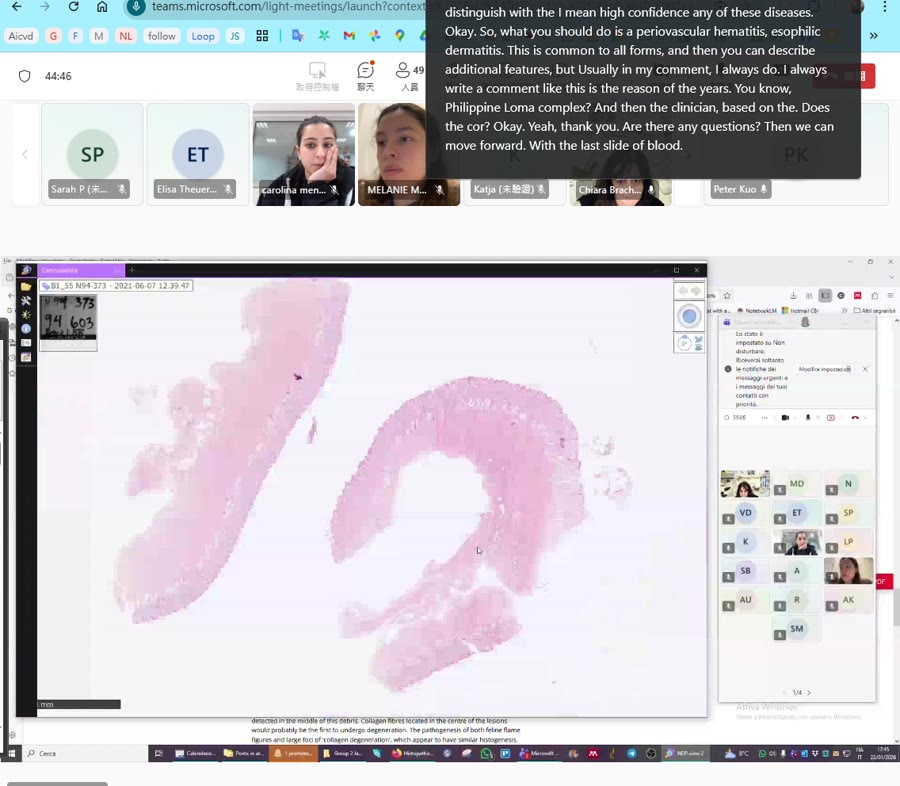
Thank you. Okay. So I believe this is a bovine, herdskin of a bovine. Yep. Yep. Yes, you can start to So, yeah, well, I will say the obvious thing that I saw, which was this big, big parasites. Yes. This guy, big guy. Yes, yes. Which I’m not sure what it is, but he’s a big guy. Yeah. So, I mean, in terms of, let’s say, big, how do you say? chapter of parasites. Are you sorry? I mean, just see, we don’t know what it is, but if you think about the size. Yes, like a lice, I’m not sure. Yeah, yeah, it is a lice. Okay, okay. But again, the same as Melanie, like she asked, how much do we need to describe these parasites? It’s… Actually, I don’t know, but I think at least the, let’s say, the major component of the body of a life, like the kitten, the muscular layers. Again, here we have gastrointestinal or usually what you see in a parasite, in a metazone parasite, it’s usually tract of gastrointestinal or reproductive apparatus. And I think this again is some kind of pieces or, let’s say, some material. Okay. Okay. Okay. Okay, perfect. So lice on a bovine. And then other things. do you have to know the name? Do we have to know the names? I don’t know. Well, I don’t know these. I don’t know the name of the lice. I have no idea. We should ask, I don’t know who we should ask, actually. Third year, maybe do you know the names of the lice? We should know there is… We should, yeah, I guess. We should. Okay. I don’t know. Okay, I didn’t study. I have no idea the names of… Yeah, I think there are different types of lives. I don’t know. I personally hate the parasite. Exactly. I think it depends on the way of feeding. So basically if they are feeding with scales, with cellular debris, and with hair, or if they are hematophagus. And depending on this, they have, of course, they have different names. They belong to different species. I have doubts that we can tell by history of the difference between the two main type of flies. Okay, yeah, yeah, probably. I think these two looks very superficial. I mean, it’s just sitting on top, so we don’t know. Yeah. But I think we cannot tell from the histology. Okay. So you presume a bovikola bovis maybe. The bovicole voice is one of these keratin eating stuff, right? It’s not a ematogenous. No, emacs. What did you say? Oh my God. and matogenous blood feeding yes yeah okay and that’s it and then well diffuse even though it’s a bovine a root basket weave let’s but i don’t i’m not sure let’s see it again yeah i don’t i mean to me looks a little bit more lamellar but okay yeah I mean I think we can discuss a moderate typical malibal malibalibalibalibal and the mild interstitial infiltrate or peri vascular maybe perivascular to interstition yes infiltrate and I’m not sure what it is the cells oh my god what are these yeah I have to say that immediately below the the lice, the scanning is not very good. But yeah, you can get that there are some eucinophil around, but they are not very many, eh? I mean, this is not severe peri-vascular dermatitis. This is a little bit more than normal dermis. But you see, for example, here, here, these are all eosinophilts. Okay, okay. Okay. Okay. Okay. That’s it, I think, no? Yeah, I think the hypertherosis, the peri vascular inflammation, mainly, eosinophilic, and the parasite. I don’t have anything else to say, too hard. Okay. Okay. So what would be the more? I mean, here for example, if you took, if you take by comparison this piece of skin that is more or less fine, you see that there is these inflammation or this perivascular cells, but in the other piece where the lice is is more prominent. So you have more inflammatory cells. but even in the other piece is not so severe. Okay? Okay, so not severe. Yeah. Moderate. Yeah, mild to moderate. Malt to moderate. Chronic, still, even though we have eosinophiles, because we have a perplasia, diffuse perivascular to interstitial isinophilic dermatitis. With the parasite, how can I, with the lice figure, like, with the intelligent lice. Like, with the, with intelligent lice. Nice, okay. And maybe we should also put the hyperclerosis on the more. Okay. I don’t know. Okay. Yeah. Okay. add to that hypercertosis. And what would be the name of the disease? Pedicolosis. I think, okay, perfect. Okay, and then we are nice and we want to ask you the name of the life. Thank you very much. At psychology. I don’t know. I don’t know. We are free. Okay, so I think today maybe we are very on time and maybe a little bit early. So if you have some questions or if you want to see something specific or otherwise. Yeah, we had to check the screenshots from Nicole in the discussion. Yes. I would like to ask a question. if it’s possible. Yeah, of course. Besides the size of the parasite and the fact that it’s on the skin, can we use other elements to presume or to be sure, in fact, that it’s a lice? The right answer is yes for sure, because I am sure that there are differential features.
🖼️ [00:52:14]

between the different parasites. However, the honest answer is that I am not able to tell you because I don’t know. I used to know. When I sat the board, I used to know. But I sat the board 20 years ago, so I forgot everything. What I can do is I can try to find if I still have somewhere the notes that I had done, with all the parasite. I don’t know if I find them. Thank you. Okay. Okay, thank you very much. No, no problem. I’m sorry, I cannot help you. So do you see still my screen, right? Yes. Oh my god, I realized that we forgot to record the session. I am sorry. We also forgot to remind you. Yeah, sorry. We try. It’s still new. No, I think, I mean, I will remember at the end of the year. It takes me one year to put it in this empty head that I have to record it. Sorry, I’m so sorry. So, this exactly screenshot is just the kind of normal artifact. that you have when you have some retraction of the nuclei. And this is often happening, especially in the superficial layer, okay? So, and as you can see here, it’s quite homogeneous all around the nucleus. It’s not really, let’s say, asymmetrical. So I would not think that this is a very, um, uh, a real, let’s say, perinuclear fagul. I would just say that this is a little bit of retraction, normal artifact due to fixation. Oh. Okay. The second pictures she put in, I think it really looks a bit more like the
🖼️ [00:54:45]
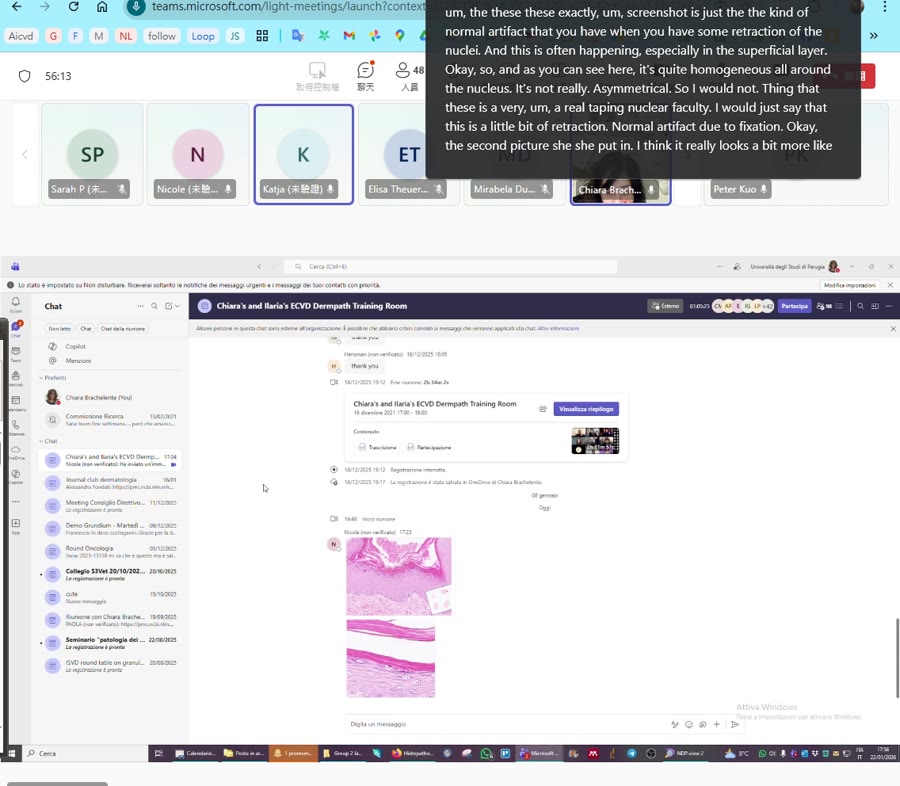
vacualization, but it’s probably too less. Yes. Again, this is just one. I agree it could be. It could be. I mean, if you look at this picture and the picture of the paper from Elizabeth, it could look like. However, in the, and we have, we should check if in the paper of Elizabeth, the keratinocytes is still viable. Because here I have the impression that the nucleus is gone. Whereas I, if I remember correctly, the picture that they are showing, there is the nucleus. that is viable and then there is this perinuclear hollow
🖼️ [00:55:32]

wait a second we look at it together we have time you know what’s the first one yes here that vaculated keratinocytes in the stratumcona no but there was another one there was another one with the color
🖼️ [00:56:28]

🖼️ [00:56:34]

pictures. Yeah, this one is no.
🖼️ [00:56:52]
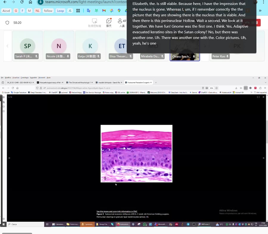
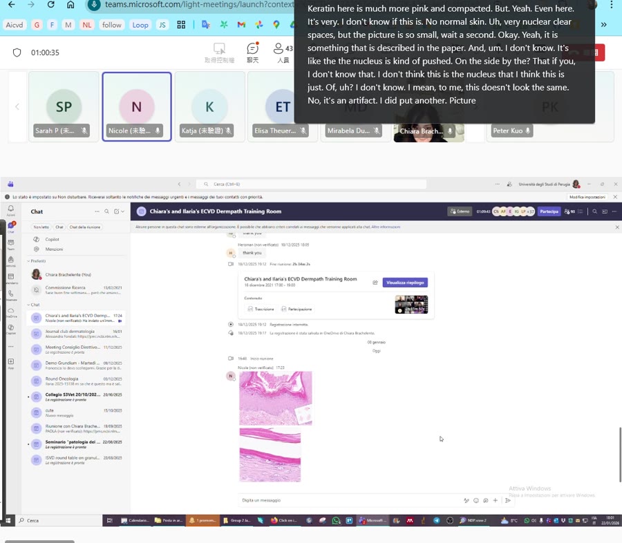
Okay, this is the autosolar resettificiosis of the American wood dog. But you see, I mean, the nucleus is still okay and there is this vacuum. I don’t know. I don’t know. And the carrot in here is much more pink and compactive. Yeah. But yeah, hold on the other,
🖼️ [00:57:22]

yeah, even here it’s very, I don’t know if this is,
🖼️ [00:57:47]

no, normal skin, very nuclear clear spaces, but the picture is so small, wait a second,
🖼️ [00:58:01]

okay, so, yeah, it is something that it described in the paper. and I don’t know, it’s like the nucleus is kind of pushed on the side by the, that if you, I don’t know, I don’t think this is the nucleus.
🖼️ [00:58:44]
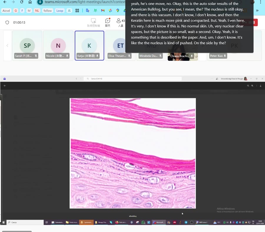
I think this is just a stain of, I don’t know, I mean, to me this doesn’t look the same. No, it’s an artifact. I did put another picture.
🖼️ [00:59:06]
This is from Elizabeth Mulden. It’s a color picture, but it’s also really small. But I just see these two pictures. Oh, wait a minute. Me too. And now? Okay, now it’s coming.
🖼️ [00:59:27]

Yeah. So for me it looks the same.
🖼️ [00:59:34]
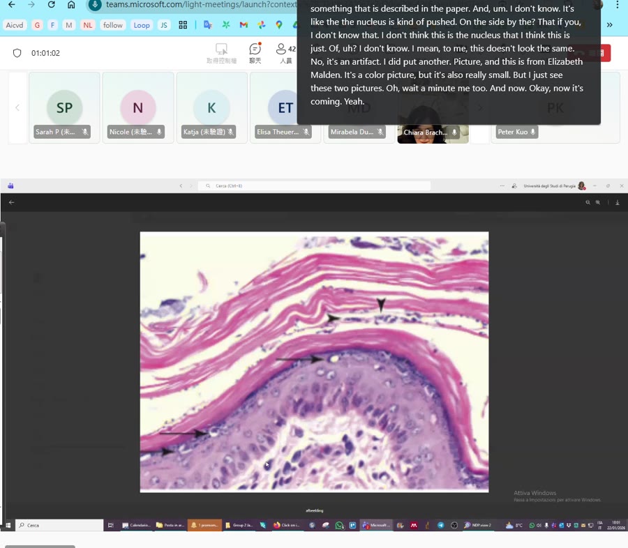
Yeah. I don’t know. It’s really hard to tell. Yeah, it’s hard to tell. And maybe here you have more of these. I don’t know. I think… And when you look at the hypercarotor, so that was… That confused me, because for me, it really looks alike. But… No, I see what you mean. Sometimes, if you look at the single detail, you cannot really tell the difference. And I agree, I told you that the hypercalatosis should be more pink, more compact, but here, for example, it’s more lamella, it’s not very compact, and of course you need the clinical picture together with it. But in general, I think that this, I mean, if you just have one of these, and in the Malacetia case that we had, there were not many, I have to say, right? Yeah, you agree. Yeah. And here you see the arrows, but is that also malicegia on this picture? I think so. Where did you take the picture from? This is from the… there was a book with a chapter, Elizabeth Mulden wrote. And can you check the figure legend? I think these are malacetia, right? It is… And I first have to find the book and then I have to find the chapter, so I guess it will take some time, sorry. Aha, no, I thought that you took the screenshot now. I mean, just now. No, no, no, I had it in my notes. It’s from M. Peters. Okay, so it’s the Jabben Kennedy, I think.
🖼️ [01:01:44]
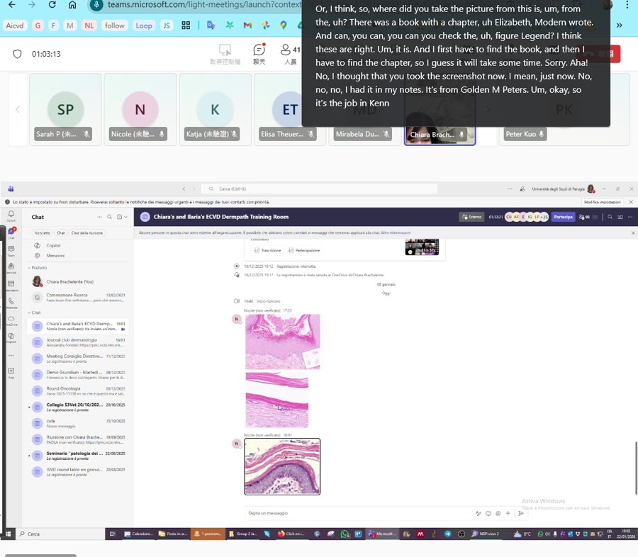
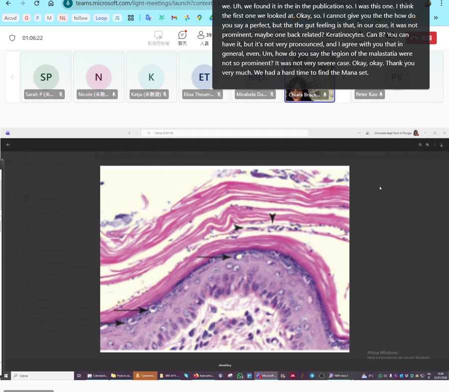
Yeah, it is. For the second, I have the old one, I mean, the one that is out now, I don’t have it. My computer is leaving me. So, it’s page 554, 555, this. If you tell us what it’s written, I don’t need to. I’m almost there, I’m almost there. So if you have any, here I have it. It’s an American, it’s American Bodok ichthyosis. Note the expansion of the stratum corneum by diffuse lameler ortho-keratotic hyperkeratosis with proliferation of melasegia. Gradually can’t exercise have perinuclear clear spaces.
🖼️ [01:03:24]

Yeah. And in these picture there are quite many of these characteristics. that have this perinuclear aloe. Mm-hmm. And this is an American bulldog. It’s not a golden retriever. I don’t know if in the American bulldog, it’s even more pronounced is perinuclear aloe, but I remember that it described even in the golden retriever, and we found it in the publication. So it was this one, I think,
🖼️ [01:04:00]
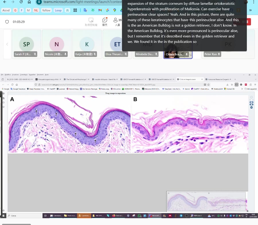
the first one we looked at. looked at. Okay, so I cannot give you the, how do you say, perfect, but the gut feeling is that in our case, it was not prominent. Maybe one vacillated keratinocytes can be, you can have it, but it’s not very pronounced. And I agree with you that, in general,
🖼️ [01:04:34]
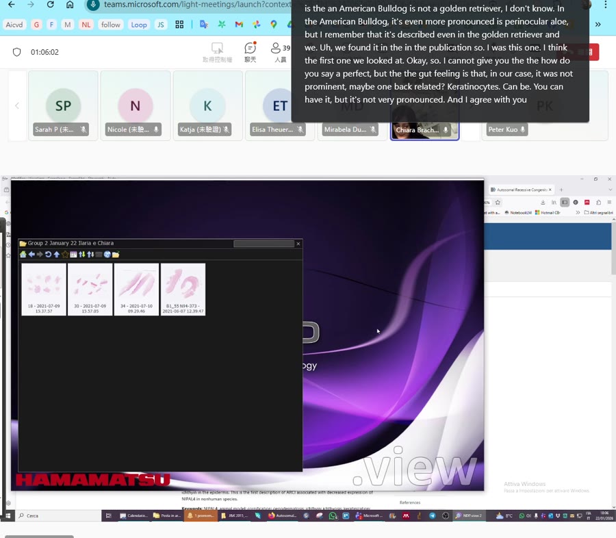
even the how do you say the lesion of the malacetia were not so prominent it was not a very severe case okay okay thank you very much we had a hard time to find the malasteti okay so I think I
🖼️ [01:04:54]
can stop sharing the screen and I can say bye okay
🖼️ [01:05:02]

Thank you. Thank you very much, Kiaram. Thank you. Bye. Bye. Bye. Bye. Thank you very much.