2025.12.19 ECVD HistoRound(自動整理)
由
錄製_2025_12_19_00_01_54_743.mp4自動產生:faster-whisper 逐字稿 + ffmpeg 切片截圖。 ⚠️ 這是機器自動整理的原始版(英文逐字稿可能有小錯、切片圖是自動抽的)。看過後再自己濃縮成重點、補中文。
- 已人工校對/濃縮
🧩 內容(切片 + 逐字稿,依時間交錯)
Okay, so let’s start. Who would like to volunteer? We have four cases today. I think that probably we will manage to do three, two or three. It depends on you. So is there anyone who would like to try?
🖼️ [00:00:17]

And start, if you want, I have the slider B585. Okay. Do you want I share my screen? Do you want, as you prefer, you can share your screen, I can share the screen.
🖼️ [00:00:29]

But it’s better if you share your screen because you are three residents, so you should be able to navigate. So I will share it just two minutes. I open this, yes, like that, and then share the screen.
🖼️ [00:00:54]

Okay, so now you see my screen.
🖼️ [00:01:01]
Okay.
🖼️ [00:01:07]
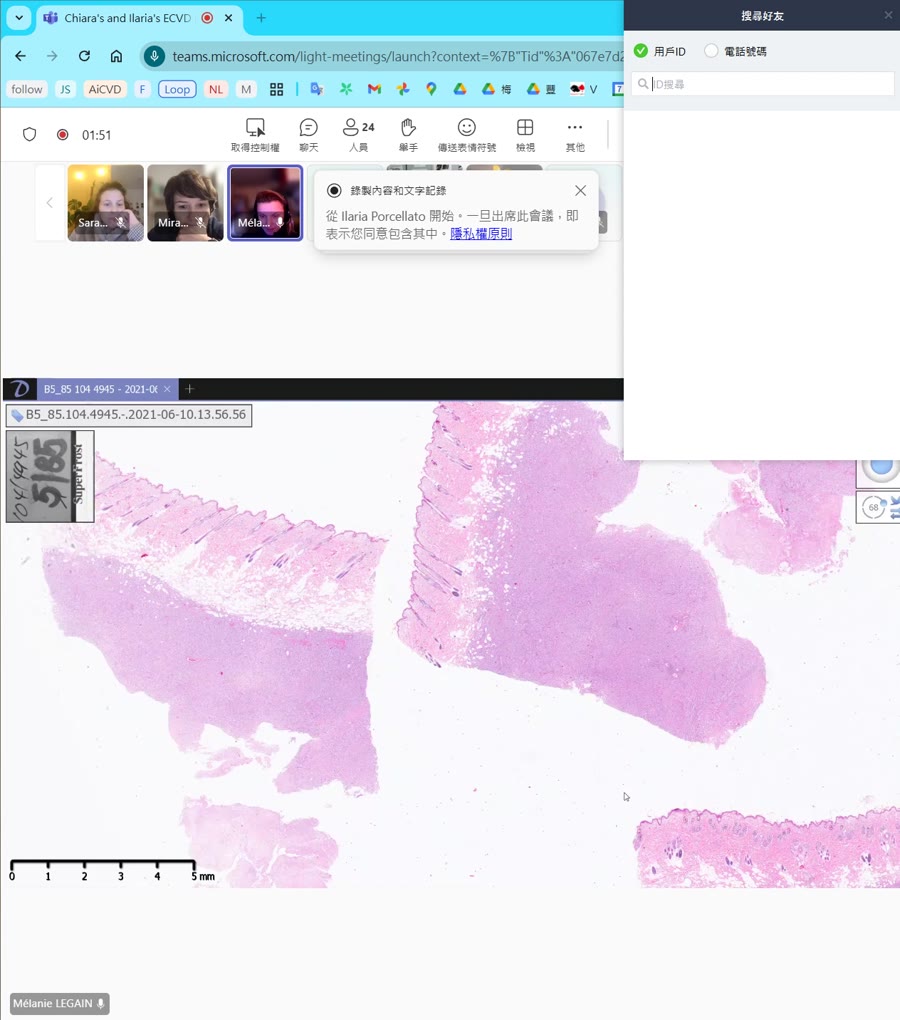
Maybe I just put two, because I just want to see what I have prepared.
🖼️ [00:01:13]
Okay. Perfect. So we are in skin two more. So I use the seven sentence type description. We have, I find whale delimitated, densely cellular, nodular, and expansively growing. It is unencapsulated. We can see with the border of the tumor. Skin tumor, focally expanding to the whole dermis and also to the subcutis. Concerning the margins, I think it is infiltrative because we can’t see in the back of the portion. I think we have cells that are again in the tissue like here. The cells are organized in sheets. We have a scant fibrovascular trauma with foci of nodular, inflammatory infiltrate like I can show you here composed by lymphocytes and concerning the cells now they are 10 to 20 maybe sometimes more than 30 micrometer one cells with cell borders that in the slide are not very well delimitated sometimes yes but sometimes not like here The cytoplasm is large, pale, basophilic and homogeneous. The nucleus is most of the time unique rounds to ovoid, because here very ovoid, here more round and eccentric. The chromatid, here is not a good example, but sometimes we are very typical chromatin, I think, like here. So it is the chromatin is coarse, clamped and arranged in cartwheel. Concerning the nucleus, they are nucleus. One, two, three, maybe more sometimes. And the anisocaryosis and these cytosis are very, very high, and we have giant, bizarre cells. No special features. Around five meters per field at 400 multiplication, I don’t know the name. Highland field. Thank you. And some atypical mitosis. Few amorrhages, like in the superficial, near to the superficial dermis. I don’t remember where, maybe here, not emmerage here. Like here, but they are some larger area. So for the morph, I think it’s a plasma cell tumor. And I know there is subtype, but I am unable to find the subtype. A subtype for plasma cell too? I see an article with a description of subtype of plasma cell tumor.
🖼️ [00:04:47]
the article talk about fibroid. Oh, okay, okay, yes, there are some articles that they try to, like, give some subtypes of plasma cell tumors, but as pathologists, we say plasma cell tumors. Okay, perfect. Because I passed maybe half an hour at France, I don’t know, but it doesn’t make this. No, and it doesn’t even make sense because there is no like a prognostic significance or I don’t know, maybe, it does not have anything. Okay. So, okay, did you describe something else in the slide? No. Okay. Okay. So is there anyone else who has something to add, something to discuss with Melanie? Yeah, I have a question. If we are talking about a plasma cytoma, plasma cell tumor, do we need to expect these Rossell’s bodies all the time? Do we have it to, sorry I didn’t get you? The Russell, do you have a, what’s the name to say? The Russell’s bodies? The Russell’s bodies. Yes, the Russell’s bodies. When you, So, so, normally, I mean, in plasma cell tumors, Russell bodies are not always present. They are almost always present or they are typical in, for instance, in stomatitis in cats or, for instance, in plasma cell podermatitis in cats. So there you can see really a lot of russaractics. body within mot cells. So plasma cells, it’s basically the same. I mean, mod cells are the cells that contain russed bodies and they are quite a striking features of plasmacetic inflammation, particularly in cats, but also in dogs you can see that. In plasma cell tumors they are not so frequent, not so prominent, what is more prominent in plasma cell tumor is the presence of binucleated, multinucleated cells, and as Melanie was correctly pointing out, also the cartwheeled arrangement of chromatin. But Melanie was consistent in her description because she went for sheets when she was describing the arrangement of cells. and so she went for a round cell tumor, which is in this case plasma cell tumor. But do you all agree? Obviously, Melanie, we are just discussing together. I’m not saying, oh, and there’s a problem. It’s just too. Do you all agree with the fact that we are facing round cell tumor here? It is. Oh, hello. Yep. I actually thought it was misanhumored cell. Okay, okay. So this was pretty hard to tell. But if you go down, that was perfect, Melanie, this area was perfect, this area that they were going like to zoom in. And if you just go down and take a look closer at the slide, you will see that the cells are not arranged in sheets. They are arranged in bundles. And you can see that the cells are like stretched, long. In some areas it is more prominent in some areas is less prominent because also it also depends if the The bundle is cut longitudinally or transverse in a transverse section But if you just take a look and see the cells here are Are spindle and maybe Melanie. I can share my screen so I can show what I mean Okay, but thank you for your description which was really nice
🖼️ [00:09:05]
and clean. Where did I lost it? Okay. Here. Okay, here. Share. Here it goes. You can see it right now. Okay. So if you go down here, you can see that the cells are pretty stretched. Okay. I agree with you in this area. They look rounded. Okay, but I mean is not It’s not like they are really really round cells in this case They are more mesenchymal and I agree with the I agree with you when you say that they With I don’t know who said that but yes they are they look mesanquimal to me I agree with totally agree with the description you did so you said that the cell market were sometimes visible, sometimes they were fading. And that’s another point. In plasma cells and in ron cell tumor in general, the nucleus is round, sorry, the cytoplasm has well-defined margins. And you can see, like, you can tell where is exactly the end of a cell and the beginning of the next cell close. In this case, it’s difficult to see, okay? Do you know what I mean? Do you agree with me or is it still a bit foggy? Like, okay. I suppose that it was because the malignity of the tumor was high, that we can’t sometimes discern the border of the cells. But yes, I agree. Yes, but also with plasma cells, plasma cell tumors, I mean, even though they can get quite atypical and anisocytosis, and an isocaryosis can be quite prominent, but you will never get these spindle cells. These are frankly spindled, okay? So if you have some doubts and you see some areas where they look rounded, like in this case, just take some moment and, like, go round in your slide, in your image, whatever you are provided with, and make sure that is not a transverse section of your cells because that is the point. Here they are cut like transversely and here they are cut longitudinally. So you can see the whole length of the cell. I totally agree with you on the fact that you have dramatic anisocytosis, anisocytosis and anisocaryosis and the fact that there are really dozens of mitosis and they are mostly atypical mitosis, okay? So you have like chromatin, which is, is distributing really randomly, as you can see here probably. And also here, I mean, you cannot see like a nice equatorial plate of the mitosis. You know what I mean, what I’m talking about? Okay, so here again. Another thing that I would probably have focused on is something that is in the cytoplasm. and here I think you can see it in the cytoplasma of the cells you said it was both of phylic and homogeneous melania yeah yeah okay do you are you still convinced it’s all homogeneous no no but it’s fine I mean it’s totally normal okay so what you what you see here like yours yeah there are optical empty vacuoles and sometimes this vacuum vacuum The vacuole also is like, as you can see here, is against the nucleus and sometimes it can deform also the nucleus membrane. These are subtle things obviously, but it’s just to give you an idea of what can happen also in a neoplastic process. But there are these vacuoles, these empty vacuoles. And when you see empty vacuels on H&E slide, what do you think about? about macrophages and eustocytes and so if we are in mesenchymol tumor so it is i don’t know the name in english sarcoma with macrophages is you sitic sarcoma oh there yes yes okay i i mean i’m i’m totally agree with you this might be a differential for this this case this was not the case of an isheucytic sarcoma because usually in but i know it’s it’s difficult when they are so undifferentiated is not so so easy but in cytology you can see the backwoles in hystisiotic sarcomas in hystisiotic sarcomas is difficult to see such so large empty vacuoles these vacuels are lipids okay so when you see this vacuals pretty large rounded that like sorry there is someone with the microphone open but okay okay and so these vacuels are generally sorry I was I thought I switched off the the cold I’ve generally nipids so when you see these things these are lipids also in liver when you see liver with this kind of vacuals is usually a stertotic liver, okay? So I thought that would be the rest of the paniculus because this tumor invaded the paniculus and this is the rest of it. This is a nice idea, this is a nice idea, but and I agree with you in these areas. Okay, so here you have these are adipocytes probably or lipid that are entrapped from the like the paniculus that is infiltrated here but in this case if you go down and just take a look you will see that they are inside the cells so either they are phagocytose by the cells or they are produced by the cells okay so when you think about cells that produce or store better lip it what do you think about Glens. Sorry. Sebaceous glands? Sebaceous glands? Yes, it could be. But I heard something that I like more. We are in the paniculus. Eiposarcoma. A liposarcoma. Okay. So this is the case of liposarcoma. I totally understand that this is, it might be difficult. But the important thing is that you here get the fact that this a mesanimal tumor, that this is a spindle cell tumor, okay? Then, I mean, if you, the diagnostic key for a liposarcoma, once you see that, you will always remember that, is that when you go down, you see and you find these cells with these rounded empty vacuoles. And usually you recognize a liposarcoma also from a low magnification because you can find these like more vacuble, raffle filled areas I would say okay so you can see here these are lipids and these are inside the neoplastic cells okay so just take a look if you have like and you see it’s a spindle cell tumor and I mean in this case you are also in the panneculus so it’s the ideal place for a liposarcoma to arise and if you see also from a low magnification that you have these areas which are clear, like more clear, just go down and check. If you see and you find that there are these empty vacuoles, then it might, it could be your first differential, okay? But I totally agree that in this case, if you said that this could be is you see the sarcoma, I mean, as a pathologist, probably I would say meh, but I mean, the morphology of the cells, because I see the vacuoles and sometimes, I mean, the vacuels are pretty, pathognomonic, I would say, but in this case it could be because there are multi-nucleated cells because you have different shaped cells and also isisiotic sarcomas can show also sometimes this spindle appearance of cells. This, I mean, if you, I mean, if you weren’t visualizing the, the, the, the, the, the the empty backwoods here this could have been a hysthydeocytes and i would have nothing to um let’s say to to to comment to that but in this case you have also other histological features to consider okay okay if it’s clear do you have any other kind of questions? Okay. Was it hard for you this case or I mean apart for Melanie but I mean you spotted this was a tumor and you described it correctly I mean the structure of the description was perfect so don’t worry about like in finalizing the your your comprehension of what you are seeing. So it’s totally normal. Okay, so can we on with the next? I can continue with the next one. I have the B775. I will try to share my screen. Yes, I just stop my, okay. So your name is very, is very long you are i’mara okay i’ma oh wait i think i need to wait a second okay i need to leave the call and then try again okay no i will tell no to start here okay can you see my screen probably no no okay but i’mana i can i can share it for you and you can tell me where to to go so if you want to okay yes i think it’s better because then we need to take I know. Okay. Okay. Just a second and please repeat the code. B775. Yes. Okay. Okay. So first I will go with an end, so we need to wait. Yes. Just screen. Okay. Okay. Now you should see it. Yes. So yes, we see a large mass or neoplasma. So I wanted at the beginning to show, okay, where I’m looking now, I’m looking to the hair skin. And then I found one small area of hair skin. Yes, there. And then we can see that the sebaceous glands are uniform, are nice. So I thought this is a cat. So we’re talking about hair skin from a cat. And then we have a nodular. non-encapsulated, expensive, poorly demarcated dermal neoplasma, so it’s all the dermis is affected. Yes. With infiltrated margin, so I cannot define if all the neoplasma of their tumor is more excisive. The nodule is composed of a densely packet spindle cells. and half a I cannot pronounce this word a hazardly yeah thank you arrange and with vascular space so we have there is vascular structures in between the cells or then the spindle cells and there is also if we look more close we have also a moderate infiltration of mass cells and this is two spects because it’s a cat. And the cells are elongated with no defined borders. There is an homogeneous eosinophilic, no, no cellophilic cytoplasma. The nucleus is oval to elongated. So we have run also oval to elongated in nucleus. And the center are central and also Pignotic in some of the cases or in some of the nuclear. Yes. And we have also present of as one central nucleoli. Nuclearolus. Nuclearolus, okay, nucleolus. And there is no other special features present. And metotic figures are few of them. I only see one or two per ten, high power field. And we also have anisocaryosis from the nucleus. Okay. Yes. And then I described the epidermis because we have also features of the epidermis. So we have a large, so we have a severe hippoblacia of the epidermis with large areas of ulceration and coagulative necrosis. And we have also multifocal areas of emirash and around this ulceration, we have also an infiltration of inflammatory cells, probably secondary, dominated by plasma cells, lymphocyte, and neutrophils. Yes, I would say that here probably they are dominated by neutrophils. There are also plasma cells and lymphocytes down here, but when you have an ulceration, you have across, in this case, you have also, I think that I saw somewhere some bacterial overgrowth. Yes, yes, obviously. Yes, and also eosineophase as well. Yeah, I forget eosineophiles. Yeah. And my diagnosis is a feline sarcoid. Okay, correct. And yes, I just wanted you to focus on, do you know, have you ever seen equine sarcoid? not life okay it’s basically it’s basically identical so histologically it’s really really the same there are some key features that you need to describe when you are facing a sarcoid and this is you correctly described the fact that there is this hyperplasia of the epidermis and this is one of the key features because when you have feline sarcoid or an equine sarcoid, the typical feature is the presence of these deep retpegs or retary ridges that grow inside, I mean, that grow deeper into the dermis within the neoplastic cells usually because neoplastic cells reach up to the growth of neoplastic cells reach up to the dermepidermal junction. The second feature that you might see is this one, probably here is you can see that. Can you see that these cells, neoplastic cells, are like arranged perpendicularly to the, yes. This is called a picket fence appearance. So they are arranged like in a palisade. They are palisading. And this is another typical features. You may not see that everywhere because, I mean, for instance, if you go here, you cannot see it. But then here you can like recognize it a bit and here again is coming out again. How’s the name again, sorry? Picket fans. I write it down in the, because I don’t know if my pronunciation is good, but it’s like the, and I don’t know how to explain in English so I will write down as soon as I stop sharing. So okay. Can you, can you, can you, sorry? I wanted to know if that’s typical for sarcomas. This is typical for sarcoids. This is typical for sarcoids. So both in equine sarcoids and in feline sarcoids, red pegs, and the presence of these picket fence are like key features. And I mean, picket fence, it depends on how the tumor is section. Sometimes you might not see that, but usually when they provide you a picture during an exam, they will find, I mean, just give you an area where you have that because it’s quite a diagnostic feature of these kind of lesions. you correctly described the fact that the neoplastic cells are are fastedly arranged and there is something that I think you should have described together with the neoplastic cells it’s the collagenous matrix there’s a lot of collagen here so they are embedded in abundant the eosophilic, finally fibriilar material, and in brackets you might write mature collagen, okay? And another thing that might be like when you said that there are vessels, I would write small caliber vessels or small capillaries or capillaries because they are usually very, very small vessels, Okay. Or you can write down in this case sometimes they are also congested and particularly linear areas of superficial ulceration. Okay. Okay. I have a few questions. Yes, yes. I thought it could be a fibroma or fibrosacoma. Would that be a differential at least? Absolutely. Absolutely, it might be a differential, but here the key feature is this one. I mean, the presence of these retipags, this like proliferation of spindle cells that reaches up to the derm epidermal junction is like a feature that is quite typical in these kind of tumors. And also here you can see that usually sarcoids and sarcoids in both cats and in the horses, you just make the diagnosis from this magnification because you see these ones. Okay. I actually thought that because you have this really, how is it called, compact hyperkeratosis there? Yeah. Yeah. And I thought that on the other edge, on this really big one, that comes down. This one? Yeah? No, no. No, on the other, no, on the left one. Yeah, yeah, okay, okay, yeah. I thought that would be a part of the paw pad, and I thought that the papillated dermis would be a pulpit. Okay, okay. And the tumor destroyed all the porpet. I mean, it could be a popad, but usually when you have this kind of growth in a in a pop-ed, then the epidermis like stretches out. In this case, there is still some proliferation of the epidermis. And then when they create these retipegs, this like in growth within the tumor is like the typical features. And I mean, it’s something that really helps you. It’s a key diagnostic feature, basically. I understand that the fact that you thought it could be a popad, and I agree with you, it could totally be a popad, might be misleading. But once you recognize this kind of pattern of growth of the epidermis, and then you see that there are these spindle cells, and usually they are not very atypical, they are like, and so I totally, and again, you are right. If you see just this field, it could totally be a fibroma, a well-differentiated fibrosarcoma. It totally, but you have to consider also what’s happening in the in the epidermis and in the dermopidermal junction because it’s a, as I said, it’s a key feature. Okay. Any other questions? Thanks. You’re welcome. Yes, can you just please show again the the picket fence yeah please yes I think that a pretty good example might be like here okay so see that there are this like okay okay they are like palisading around it’s not it’s not like nothing like a palisading that you might see in a peripheral nerve should do more for instance because it’s way more like not so orderly disposed, but as you can see here, I mean, these cells are perpendicularly arranged if you consider the epidermis, okay? So that’s basically it also here. I mean, you see that there is like this pattern going like vertically from the, from the epidermis. And so, yeah, this is it. and so if there was I mean do you know the etiology of this kind of lesion some bovine papillomavirus infections yes but which one one and two I don’t know it was I mean some years ago it was written that it was bovine poppy Loma virus one I think and but then in 2015 I think they sequenced the papillamavirus within this lesion. It’s a bovine papillomavirus, but it’s bovine papillama virus type 14. So it’s very similar. It’s in the same played from bovine papilloma one, papillam virus one, two and 13, if I’m correct, but it’s number 14. And it has been, it seems like it’s specific to this kind of lesion. Okay. Okay. So it’s probably the only example or one of the few examples known at the moment of cross-react, sorry, of cross-species infection of papillomavirus, okay? Because papillomavirus are species-specific. So if it’s a bovine papillomavirus, it can infect only bovines. If it’s a feline papillomavirus, it infects only feline. And this bovine papilloma virus is an exception. I think there is some exception also in ovines, but I studied that, but I don’t remember, I don’t want to be misleading. Still, this is the most known, well-known example of species. Okay, next one. Is there anyone who would like to volunteer? I can continue with B16. Okay, thank you. Mirabella, right? Yes. I remember correct. I would appreciate if you can drive because I don’t have the slides down on my computer. Okay, okay, okay. No worries. Let me find out where. So you said B-877, B-16. Okay. Okay. So here we go. Oh, I love this slide. Okay.
🖼️ [00:35:45]
So from this view, you can already see that the important things are happening in the epidermis, in the superficial and eventually in the middermis. I can presume that from clinical point of view I have maybe a plaque like lesion or a nodule. type of lesion. I can see at least on the sample that it’s in the lower part of the screen, yes, that one, that I have some like a nodular pattern of aggregates of cells that probably are organized around the annexe. I tried to see if it’s hair skin or not. I can see some hairs. usually at the margin of the sample. So I presume that maybe it’s a sparsely-haired skin or it’s like mucocutaneous junction, maybe. And seeing the fact that I don’t have an abundant paniculus, maybe it was like the face, the areas where they took the sample. Yes, and I just wanted to show you, Mirabella, that in this area, you said that it’s scarcely, like sparsely hair-de-skinned, but in this case, these areas, if you go down and take a look, there are a lot of hair around this inside also, okay? So probably you are on the face and probably, there were quite a lot of hair. But they are not anymore. Yeah, okay. Okay. Here the pattern is quite like suggestive of it. Okay. I tried to find some sebaceous glands to see if I can identify the species. And honestly, it was not very successful. Okay, okay. So I did not manage to find the, to find sebaceous glands. Okay, I think I can tell you this probably from what I can remember it was a dog. Yes, it’s correct. It’s dog. And yes, you are also correct because I was thinking that when I was seeing from the distance. This is a Mukukutanis Junction. This sample is taken from Okutans junction. Here you have hair. Here you don’t. Okay. Yes. So. Probably also here. it’s like on the and flip so this is the the mucosal side and this is the mucosal side okay we can already tell that the in some areas the the epidermis it’s gone so we have erosions and ulcers on the surface of our of our sample Then I tried to check what type of cell are predominantly present in those aggregates of cells. And I’ve noticed that we have many lymphocytes, but there are also some other cells like histocytes, like plasma cells, like neutrophils in some areas. epidermis it’s also infiltrated by these lymphocytes which are sometimes really like dispersed in the in the epidermis but sometimes they are also organized as aggregates as small groups of of cells which I associated considering the the aspect of the sample and also the team of our meeting today with Potier Obsesses. I’ve also noticed that in some areas we can observe like neptidermal cleavage, which is multifocally and sometimes important. Yes. Yes, here probably something, yeah. And also here. Also there. Yes, I checked both. both yes yes yes here there is ulceration too yeah yes there is also a focal crust but I don’t think that it has importance in the diagnosis but I I described it and I I’ve noticed it also considering the the behavior of the cells I would say and and the morphology of the cells, which I suspect they are tumoural because they are quite different in presentation. They may have different sizes, starting from small size. And there is someone with the microphone turned on and, okay, thank you. Sometimes they are quite small, but sometimes I, sometimes I got the impression that they become quite big in size. I don’t know. So how would you define this, the fact that it could be smaller or larger? How would you? I can define it as heterogeneous population and I would say anisocytosis. Okay, perfect. So you just, you can like summarize all of this different. I mean this difference and say that there are there is mild, moderate or severe depends anisocytosis and anisocaryosis. You can also split and say that there are, there is severe anisocaryosis and moderate anisocytosis. Okay. I don’t have experience, but for me it was impressive. I was not, sometimes I asked myself if there is the same type of cell due to the big differences in appearance. So for me, it was, let’s say, at least moderate and maybe even severe. It’s severe. It’s severe. I’m not sure if it is correct or not. I think it is important to mention also that the adnexia, like the hair follicle walls when they are visible, but also the, let’s say, the ancient sebaceous glands, because they are no longer. present are completely infiltrated by the tumoral cells. Yes, I think that here you can say that the follicular wall is completely effaced by the presence of neoplastic cells. I mean, here there was the hair follicle wall, the epithelium of the hair follicle, and what is remaining here is just the hair shaft, okay? And here again… Here is the same, yes. Yeah, these are epithelial cells, the remaining epithelial cells from the hair follicle, and these are neoplastic cells that completely effaced and obscured the hair follicle. Here, probably, you can see that there’s like a novel shape that could be like not so transverse, but a diagonal section of an air follicle, which is completely gone again, okay? And I don’t remember, but I think it’s easy to find. There was also a hair follicle that was cut transversely, longitudinally, and we can see the cells that are invading. I think maybe, yes, this other one, and it’s more in the superior part of the sample. It’s quite nice, because we can see actually the cells that are infiltrating the… Okay, so again here but it’s completely lost. This one, yes. This one? Yes, yes. Okay, here you have a lot of alterations because the, I mean, there’s a, it’s like, you’re probably some artifacts here. I mean, there are some, some of these tumors and I would, won’t give the answer, but, okay, some of these tumors which are nice because you can, nicer, I mean, in terms of, like, histological appearance, because you can still recognize the hair follicle. In this case, it’s completely gone. Okay. Here probably there is something remaining, and yes, here probably you have some lymphocytes, yes, but not, here, not a straightforward the potrii micro abscesses for instance here so probably some single cells in infiltrating but okay I totally agree with your description I tried to to find some some mitosis I must say that I was able to find them but for me it was like not that not that many mitosis I don’t know I did not check like every part of the sample but I tried to find them and for me I would say the metotic activity is low but I’m again I’m not sure if I’m correct or not okay if you go here and just check I mean there are one two three so and yes here anyway when you have a tumor and in this case you should know that it’s a quite aggressive tumor, probably I would not seek with low, I would say medium or I would at least try to give a numerical reference. For instance, in this case, I would think I would write one to one, two, so dash three mitosis per high power field. Okay. And so for a lymphoma is not that much. I totally agree with you. Still is not like a low mitotic end. that is something that you might find, for instance, in leo myomas, or sorry, leumaiosarcomas, where you might have one mitosis per on the whole tumor, okay? Okay, so for me, if I checked and in some fields, I was not able to find any mitotic figure, and yes, there were some fields where I found three, like the one that you mentioned, and then I thought, okay, if we do the average, maybe it’s not that high, but again, it was just a guess it was not something that I… Yeah, you can also write down and it’s something that we can, we use to do sometimes when there are no gradings to, let’s say, give for a tumor, up to. So if you find us, for instance, a high power field where you have five mitosis, then in the other fields you count three, two, you can say up to five mitosis per high power field and you give somehow a range so the like the examiner in this case might see that okay you recognize the mitosis because you didn’t see there are 80 mitosis per high power field okay I’m just giving just to give an idea what you or what you say what you see okay what you saw in this case okay okay so the the morphological diagnosis would be cutaneus epithelotropic thyselymphoma yeah correct in this case probably you were correct and here is i think uh you can see that here i mean it’s a face but it’s a lip so it’s uh epitheliotropic mucucutaneus lymphoma it’s the same it’s fine you see skin so it’s fine cutaneous lymphoma, epithelotropic, and it’s a T-cell, usually they are T-cell lymphomas. Normally, what I do, because in biology you never know, is that I write down epitheliotropic lymphoma and cutaneous epithelotropic lymphoma, and if they ask you the most likely phenotype of the cells, you can say it’s usually a T-cell informer. Okay. Okay. So basically that’s it. And did you have any, I mean, is it clear for everyone? Do you have any questions? Do you have any doubts on this case? I have just a question not about the case, but about the description. Can we use the classical description? when we have to describe this type of slide, do you use the two more type description? Because for me, it’s easier to use the classical. Always use, always use the, yes, that was something that I had in my mind to tell, Mirabella, right? But I forgot. The thing is that you have to remember to, like, like use the structure description. So start with, so what I see, where it is, if it’s infiltrating or not, the cellularity, and then the arrangement of cells that in this case Melanie would be in. Ronzel. In? Yes, so they are arranged usually in. In sheets. Correct. Yes, they are in sheets. sharing sheets and describe obviously the presence or not of a stroma. And then again, you can go down and describe the cells. When you describe cells, describe the cytoplasm, the cellular border, the cytoplasmic borders, the nucleus, which is in this case rounded. And sometimes it has some indentations, which is quite nice and is, I mean, quite typical also in lymphomas. like the indentation is not so deep as in histosytomas for instance and you I mean you do not see like the kidney shape is not so common you can see it because obviously the anisocaryosis is quite prominent and the irregularity on the nucleus are are present but is not so I mean is not so indented as in histocytoma and yeah and so describe the nucleus the arrangement of the chromatine and nucleoli and stuff like that. There is a mitosis right there. Yeah, there’s a mitosis right here. And there are quite a lot here. One, two, three, four, five, yeah. And there is something else that I wanted you to notice in this field. Did we have no more stroma? Yes, this is a densely cellular tumor. and so you might have this you might write down that there are that this neoplastic cells are supported by scant fibroblastic sorry scant fibrovascular stroma because sometimes you can see that there are some small vessels here and here but it’s really really scant and in this case you can find also that there are some what are these guys i also know if it’s yeah maybe These are eosinophils, correct. And the fact that there were some usinophil infiltrating this tumor was like something a bit tricky because usinophiles are usually associated with another type of run-cell tumor. Massal tumor. And muscle tumor can show epithelotropic activity. so it can grow also there. I mean, I think that they will not give you that in an exam, but still remember. And in this case, also the cells have quite a lot of cytoplasm, so it was not so straightforward a diagnosis of lymphoma, to me at least. But still, when we get eosinophils within a lymphoma, it’s usually associated in using the fields are usually associated with the T-cell lymphomas. Okay, so in this case, it could like the two differentials, if I had only this like field to evaluate, could have been undifferenti-differentiated muscle tumor or lymphoma or lymphoma. with likely T-sat-t-fino-type. Okay. Thank you very much. You’re welcome, thank you. I have no idea how you could define these cells as lymphocytes. I thought it was histocytes actually because I, how did you do it? I think on the margin it’s more typical lymphocytes than here in really in the nucleus of the of the tumor. If you check the margins, they are more characteristic, I would say. I thought it’s infiltrated with lymphocytes because there are few normal lymphocytes. Yes, yes. But I thought it was a hystiac disease actually. This was not easy and this is why I mean we decided. to put this in because Kara and I, we are, we have fun in giving you something to, to a bone to eat, let’s say, to chew. That’s clear. Yeah, but I mean, obviously, it was not easy, but there were, there, there are some things that might give you, like the diagnosis, first of all, the presence of the potriere micro-obsesses or abscesses. I mean, usually these epithelotropic, yes, I told you, that you can have this epitheliotropic activity also or this infiltration of the epithelium also with mastels and also with histocytomas and hystiotic tumors but you don’t get this like as i can say widespread infiltration and massive infiltration of the epidermis with this kind of tumors yes you might have some some infiltration but not that widespread usually so So first thing, and another thing that helps you a lot here is also that you have severe epithelotropism and severe infiltration also of the hair follicles. I mean, there’s no way a mastal tumor or a histo-stitoma would behave in this way, okay? Because here there is a pitiliotropism is so striking that, yes, I agree with you, the morphology of cells is quite confusing, but the pattern of growth of cells is what gives you the right tip in this case. I mean, an isiotomyatoma, how would you expect to be an osteosytoma? Macroscopically. Macroscopically. Like a nodule? Yes, a nodule. And if you think about this kind of lesion, this is, as you correctly said at the beginning, this is a plaque, okay? And anisostatoma is, I mean, it’s usually a very well-defined nodule, ovalar, and usually it’s a single lesion. You would have a single nodule. In this case, you have this infiltration of the epithelium and you have the infiltration also of the hair follicle. so like it’s following really the epithelium. So remember, do not focus just on the morphological aspect, let’s say on the cytology and the morphology of the cells, because they could be tricky, they might mislead you. So always go back also to the subgross and think, how would this kind of lesion look on the, on macroscopically how would i expect it to be in this case also the fact that this is a mucocutaneous junction would have helped you i think because is histocytomas i’ve never seen them on mucocutanic functions um and so also the localization the the localization of the tumor is not typical of an histocytoma and again muscle tumor i mean you know how a muscle tumor grows, you have seen them, and this plaque-like growth is nothing like a muscle tumors. Then again, in a diagnostic environment, real-life environment, biology can happen. So you might also experience something different from the typical aspect, but usually what we give you is like difficult to to define and but there are some tips some histological like aspects or features that can help you yeah that can help you so it’s six o’clock and unfortunately I have to to go because I have another thing to do so I have another appointment but we have one last we are not meeting on January right we don’t have you don’t have the planning yet okay I’m checking it if you have it can you share maybe so sure okay there is just one question on the discussion would you do immunostochemistry if you have doubts about being a muscle tumor or to differentiate T or B in forma like C-kit or CD3 packs okay in case probably I would I would yeah I would suggest an immunistochemistry for CD3 and CD20 to confirm the the phenotype of the tumor even though as I said before epithelotropic lymph are usually CD3 positive they might be also CD20 positive but the expression of CD20 is like something that you can see in T cells. So in this case, it would still be a T cell tumor if you have them both expressed. If I do not feel so sure, probably I would ask for Toulodin blue or a Gynne-Sustain to rule out mass cells. But in this case, I mean, if you consider the whole picture and also the subgroup, gross aspect of the tumor. I think that it could be just something to let you go to sleep without any thoughts. Because sometimes, I mean, bad days happen and sometimes you do not feel confident with what you are facing. And so in that case, it’s better to do something that makes you feel more comfortable. Obviously, don’t overuse this, like in Italy, we call that the lean it’s like the rug or something like that but anything that can help you or make you feel safe at a certain point you need to to use your knowledge and but yes if you feel like this and you want to be more sure probably I would have asked just Gimsa to confirm that or I would ask Yara who is in front of my office and so we call that the shared responsibilities so when when we write down the report we like say okay i shared with you and and so i feel more confident but it’s something that might help you actually also clinically if you don’t feel confident if you have a bad day if you feel that you are struggling with something just ask some of your colleagues This is something very precious. And even after years and years of practice and of being like specialists, it will always be precious. And always remember to ask also the younger ones and the junior ones. Because sometimes they give you a hint or a different view or a different point of view of what you are facing. Okay. It’s just my suggestion, but because I know that bad days happen. Thank you. Okay. So see you and I wish you Merry Christmas and like enjoy your end of the year party all night long and see you next year and I bring also the Kiara’s best wishes for next year. Thank you. Thank you so much. It’s so nice. Thank you. Happy evening. Bye. Bye. Christmas. Christmas and New Year’s Eat.
🖼️ [01:03:56]
Bye.